


ABIMPrep

Free lesson

GFR And Renal Blood Flow

The first lesson of ABIM Nephrology Prep Course (Nephrology), free to read.

GFR And Renal Blood Flow

Course: ABIM Nephrology Prep Course (Nephrology)

Topic: 1.1.1

Difficulty: Foundational

What you'll learn

  • PHYSIOLOGY_PATHOLOGY_DIAGNOSTICS: Explain the determinants of glomerular filtration rate and renal blood flow -> renal hemodynamic reasoning
  • PHYSIOLOGY_PATHOLOGY_DIAGNOSTICS: Distinguish measured GFR, estimated GFR, renal plasma flow, and filtration fraction -> diagnostic interpretation
  • AKI_CRITICAL_CARE: Predict how afferent and efferent arteriolar changes alter GFR in common clinical settings -> hemodynamic AKI pattern recognition
  • CKD: Choose the most appropriate filtration marker and understand the major limitations of creatinine-based estimates -> CKD assessment
  • PHYSIOLOGY_PATHOLOGY_DIAGNOSTICS: Apply autoregulation principles to board-style vignettes involving NSAIDs, RAAS blockade, cirrhosis, and heart failure -> mechanism-to-management logic

Blueprint mapping

  • Domain: PHYSIOLOGY_PATHOLOGY_DIAGNOSTICS → renal hemodynamics, filtration, and clearance interpretation
  • Domain: AKI_CRITICAL_CARE → prerenal and hemodynamic kidney dysfunction mechanisms
  • Domain: CKD → kidney function assessment using serum markers and estimating equations

EST. TIME 45-60 minutes

PREREQS None; upcoming continuity: Tubular Transport Mechanisms, Urine Concentration And Dilution

OUTPUTS You should be able to choose the correct physiologic explanation for a change in GFR, distinguish true kidney dysfunction from misleading creatinine changes, and identify when altered renal perfusion vs altered filtration pressure is driving the vignette.

WHY

Why it matters

  • Board stems frequently test hemodynamic kidney injury by asking what happens to GFR after NSAIDs, ACE inhibitors, sepsis, cirrhosis, or renal artery stenosis.
  • Many management choices in nephrology depend on whether a creatinine rise reflects expected physiology, reduced perfusion, or intrinsic damage.
  • Renal blood flow and filtration fraction are the foundation for later topics in fractional excretion, tubular transport, urine concentration, and AKI classification.
  • The exam often rewards the candidate who identifies the single arteriolar or Starling-force change that resolves an otherwise noisy vignette.
KEY TERMS

Key Terms & Must-Know Facts

1.1.1.1 Key terms

  • GFR: volume of plasma filtered across glomeruli per unit time
  • Renal blood flow (RBF): blood delivered to both kidneys per minute
  • Renal plasma flow (RPF): plasma component of renal blood flow
  • Filtration fraction (FF): fraction of renal plasma flow that becomes filtrate
  • Autoregulation: maintenance of relatively stable renal blood flow and GFR across a range of perfusion pressures
  • Myogenic response: afferent arteriolar smooth muscle constricts with stretch
  • Tubuloglomerular feedback: macula densa-mediated adjustment of afferent tone and renin release
  • Net filtration pressure: balance of hydrostatic and oncotic forces across the glomerular capillary
  • Creatinine clearance: practical surrogate for GFR, slightly overestimates true GFR
  • Inulin clearance: ideal measured GFR marker
  • PAH clearance: approximates renal plasma flow
  • Effective arterial blood volume: perfusion sensed by the kidney, not identical to total body water
  • Afferent arteriole: vessel entering the glomerulus
  • Efferent arteriole: vessel leaving the glomerulus

1.1.1.2 Must-know facts

  • Kidneys receive about 20% of cardiac output.
  • Normal adult GFR is roughly 90-120 mL/min/1.73 m2, but interpretation depends on age and context.
  • Creatinine-based eGFR is less reliable in extremes of muscle mass, unstable kidney function, amputation, rhabdomyolysis, and cirrhosis.
  • ACE inhibitors and ARBs preferentially dilate the efferent arteriole.
  • NSAIDs reduce vasodilatory prostaglandins and therefore can cause afferent constriction.
  • Severe hypovolemia lowers both renal plasma flow and GFR.
  • Efferent constriction may initially preserve GFR despite reduced renal plasma flow.
  • Filtration fraction equals \( \text{GFR} / \text{RPF} \).
  • Renal plasma flow equals \( \text{RBF} \times (1 - \text{hematocrit}) \).
  • Creatinine clearance tends to overestimate GFR because creatinine is also secreted.
Parameter What it reflects Typical exam value High-yield caveat
RBF Total renal perfusion ~1.0-1.2 L/min Falls with low cardiac output or renal vasoconstriction
RPF Perfusing plasma flow ~600-700 mL/min Approximated by PAH clearance
GFR Filtered plasma per minute ~90-120 mL/min/1.73 m2 Best overall index of kidney function
FF Fraction of plasma filtered ~20% Often rises with efferent constriction
Serum creatinine Marker used to estimate GFR Context dependent Delayed and muscle-mass dependent
Cystatin C Alternative filtration marker Lab dependent Useful when creatinine is misleading
CORE

Core content

1.1.1.3 Foundational determinants: what sets GFR at the glomerulus?

A Explanation

The first board decision conflict is this: Is the kidney problem one of filtration pressure, blood flow, or marker interpretation? The single rule that resolves the first layer is that GFR depends on glomerular capillary hydrostatic pressure, Bowman space hydrostatic pressure, plasma oncotic pressure, and the filtration coefficient. In practical test language, most stems reduce to a change in afferent tone, efferent tone, plasma flow, or obstruction.

Think in Starling terms. Glomerular capillary hydrostatic pressure favors filtration. Plasma oncotic pressure opposes it and rises along the capillary as water is filtered. Bowman space hydrostatic pressure opposes filtration and increases in obstruction. The filtration coefficient reflects permeability and surface area; it falls in disease states that reduce functioning glomerular surface area.

The hemodynamic shortcut for exams: afferent dilation tends to increase both RBF and GFR; afferent constriction lowers both; efferent constriction lowers RPF and can initially maintain or raise GFR; efferent dilation tends to lower GFR. See Fig 1.

WHY THIS IS TESTED: the exam wants you to identify which single vascular or pressure change explains the creatinine change before moving to management.

Fig 1. Determinants of GFR and renal blood flow
Exam takeaway: Match the vignette to afferent tone, efferent tone, obstruction, or filtration surface loss before choosing the next step.
flowchart TD
A[Change in kidney function] --> B{What changed?}
B --> C[Afferent constriction]
B --> D[Afferent dilation]
B --> E[Efferent constriction]
B --> F[Efferent dilation]
B --> G[Obstruction / high Bowman pressure]
B --> H[Lower Kf / less filtering surface]
C --> C1[RBF down, GFR down]
D --> D1[RBF up, GFR up]
E --> E1[RPF down, FF up, GFR preserved or up early]
F --> F1[RPF up or stable, GFR down]
G --> G1[GFR down]
H --> H1[GFR down despite variable flow]
Concept flow: How to decode a GFR question
Name the variable
Decide whether the stem is testing blood flow, filtration pressure, obstruction, or the marker itself.

Choose vessel vs pressure
Afferent change alters inflow; efferent change alters outflow pressure; obstruction raises Bowman pressure.

Predict GFR direction
Use the dominant force to determine whether filtration rises, falls, or is initially preserved.

Memory anchor: A-A, E-E = Afferent controls Arrival, Efferent controls Exit pressure. If arrival is cut, both flow and filtration fall. If exit is narrowed, filtration pressure may temporarily rise even while flow falls.
Clinician Action: Diagnose whether the stem points to altered inflow, altered outflow, obstruction, or reduced filtration surface. Order serum creatinine trend, urinalysis, volume assessment, medication review, and targeted imaging if obstruction is possible. Treat the dominant hemodynamic driver rather than reacting to creatinine alone. Refer/Escalate urgently if oliguria, severe hyperkalemia, pulmonary edema, or suspected bilateral obstruction is present.

B Worked example

A 69-year-old man with ischemic cardiomyopathy, diabetes, and osteoarthritis is admitted for poor oral intake and dizziness after a viral illness. He takes lisinopril, furosemide, and daily ibuprofen. Blood pressure is 88/54 mm Hg. Creatinine rises from 1.2 to 2.1 mg/dL over 48 hours. Urinalysis is bland. A junior trainee focuses on trace proteinuria and suggests glomerulonephritis.

Identify task: explain the physiologic cause of the GFR drop. Extract key facts: hypotension, RAAS blockade, NSAID exposure, diuretic use, bland sediment. Apply rule: hypovolemia lowers renal perfusion; NSAIDs block afferent vasodilation; ACE inhibitor removes efferent support of glomerular pressure. Eliminate distractors: trace proteinuria is nonspecific; bland sediment argues against inflammatory intrinsic disease. Verify: the most likely mechanism is combined afferent constriction plus efferent dilation on a low-flow background causing marked fall in glomerular capillary pressure.

C Exam trap

Exam trap: Choosing “intrinsic glomerular disease” because the creatinine rise is large and there is some protein in the urine. WHY IT IS TEMPTING: board takers often overvalue any proteinuria. SINGLE CLUE: a bland sediment with clear hemodynamic insults points to reduced glomerular perfusion pressure, not inflammatory glomerular injury.

D Checkpoint

Question: A patient with acute bilateral ureteral obstruction develops a sudden fall in GFR. Which physiologic change most directly explains this?

  1. Increased glomerular capillary hydrostatic pressure
  2. Decreased Bowman space hydrostatic pressure
  3. Increased Bowman space hydrostatic pressure
  4. Decreased plasma oncotic pressure

Answer: C

  • A: Tempting because hydrostatic pressure is the main pro-filtration force, but obstruction lowers GFR by the opposite mechanism.
  • B: Tempting if one remembers pressure gradients loosely, but Bowman space pressure rises, not falls, in obstruction.
  • C: Correct because back pressure in Bowman space opposes filtration and abruptly lowers net filtration pressure.
  • D: Tempting because oncotic changes affect GFR, but lower plasma oncotic pressure would favor filtration rather than reduce it.
If you missed this: Review 1.1.1.1 Foundational determinants: what sets GFR at the glomerulus? — focus on the opposing forces in net filtration pressure.

1.1.1.4 Renal blood flow, renal plasma flow, and filtration fraction: the numbers behind the physiology

A Explanation

The next decision conflict is which flow variable the question is actually asking about. Many candidates mix up renal blood flow, renal plasma flow, and GFR. The single resolving rule is that RBF includes cells and plasma; RPF includes only plasma; GFR is the filtered portion of RPF. Filtration fraction ties them together: \( \text{FF} = \text{GFR} / \text{RPF} \). Renal plasma flow can be estimated from blood flow using \( \text{RPF} = \text{RBF} \times (1 - \text{hematocrit}) \).

On exams, filtration fraction is most useful qualitatively. If efferent constriction lowers renal plasma flow but preserves glomerular capillary pressure, FF often rises. If both RPF and GFR fall proportionally, FF may remain unchanged. In classic prerenal states with neurohormonal compensation, FF may increase. In ATN, autoregulation and tubular mechanisms become less reliable, so the expected prerenal pattern may disappear.

Clearance concepts are tested as mechanism questions more often than heavy calculations. Inulin is the ideal marker because it is freely filtered and neither secreted nor reabsorbed. Creatinine is the clinical surrogate because it is easy to measure, but it slightly overestimates true GFR. PAH approximates renal plasma flow because it is filtered and avidly secreted, so almost all PAH delivered to the kidney is excreted in one pass at low concentrations. See Fig 2.

WHY THIS IS TESTED: the exam wants you to recognize which parameter changes in ACE inhibitor use, volume depletion, or efferent constriction rather than getting lost in terminology.

Fig 2. Blood flow, plasma flow, GFR, and filtration fraction
Exam takeaway: Translate every renal hemodynamic vignette into RBF, RPF, GFR, and FF rather than relying on memorized drug associations alone.
flowchart LR
A[Cardiac output] --> B[Renal blood flow]
B --> C[Subtract hematocrit]
C --> D[Renal plasma flow]
D --> E[Filtered portion]
E --> F[GFR]
F --> G[Filtration fraction = GFR / RPF]
H[Inulin clearance] --> F
I[PAH clearance] --> D
J[Creatinine clearance] --> K[Approximate GFR, slight overestimate]
Fig 3. Labeled renal microcirculation and filtration forces
Exam takeaway: Visualizing afferent inflow, the glomerular tuft, and efferent outflow helps predict whether a change primarily alters flow, pressure, or both.
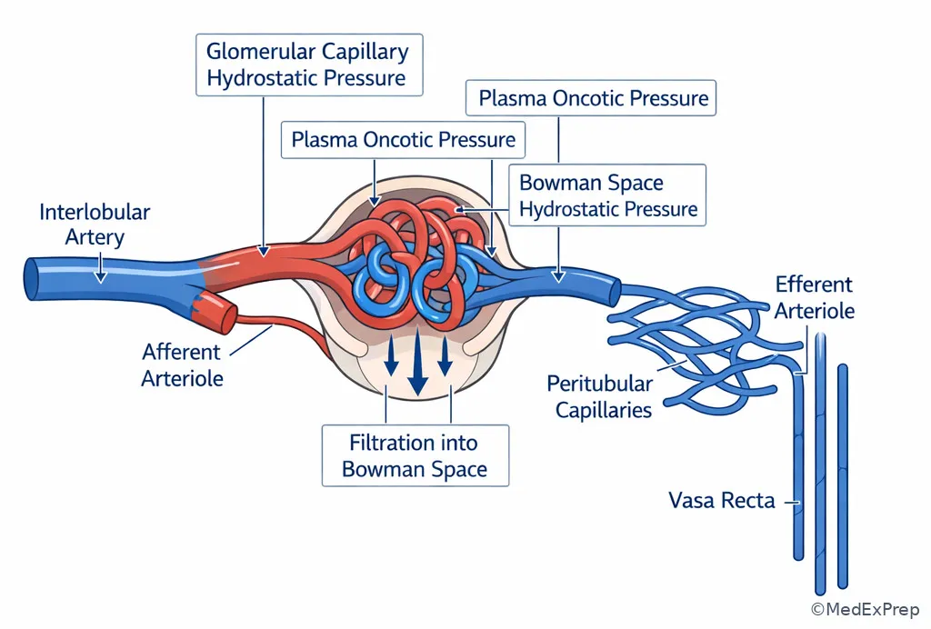
Fig 3. Labeled renal microcirculation and filtration forces
Concept flow: Which renal variable is being tested?
Start with the wording
If cells are included, think blood flow; if plasma only, think plasma flow; if filtered output, think GFR.

Ask if a ratio is implied
If the stem asks what fraction of plasma becomes filtrate, the answer is filtration fraction.

Attach the marker
Inulin measures GFR, PAH approximates RPF, and creatinine approximates GFR clinically.

High-Yield Connection: This concept is tested alongside Fractional Excretion Reasoning and Prerenal And Hemodynamic AKI. Know both the hemodynamic mechanism and the downstream urine index pattern for exam day.
Recall: This is the opening physiology topic of the course. In the upcoming topic Tubular Transport Mechanisms, these flow variables become the delivery side of transport: filtered load only makes sense after you know what was filtered in the first place.
Clinician Action: Diagnose whether the problem is reduced total perfusion, reduced plasma delivery, reduced filtration, or misleading serum-marker behavior. Order serial creatinine, urine studies, hemodynamic assessment, and consider cystatin C when creatinine is unreliable. Treat the perfusion problem or drug exposure causing the abnormality. Refer/Escalate if there is progressive AKI, hemodynamic instability, or suspected renovascular/obstructive disease.

B Worked example

A 58-year-old woman with cirrhosis, tense ascites, and type 2 diabetes is admitted with rising creatinine after large-volume paracentesis without adequate albumin replacement. Hematocrit is 30%. A question asks which renal variable is most likely increased as compensatory angiotensin II levels rise. Bilirubin is markedly elevated, which distracts the team toward intrinsic tubular injury.

Identify task: determine the hemodynamic variable changed by angiotensin II. Extract key facts: low effective arterial blood volume, RAAS activation, cirrhosis. Apply rule: angiotensin II preferentially constricts the efferent arteriole. Eliminate distractors: RPF tends to fall, not rise; total renal blood flow is not the preserved variable; GFR may be maintained transiently. Verify: the best answer is an increase in filtration fraction.

C Exam trap

Exam trap: Choosing increased renal blood flow in any state where GFR is preserved. WHY IT IS TEMPTING: candidates assume stable filtration means adequate perfusion. SINGLE CLUE: efferent constriction can preserve GFR while renal plasma flow actually falls, making filtration fraction rise.

D Checkpoint

Question: Which clearance most closely approximates renal plasma flow in standard physiology testing conditions?

  1. Inulin clearance
  2. PAH clearance
  3. Creatinine clearance
  4. Urea clearance

Answer: B

  • A: Tempting because inulin is the “ideal” marker, but it is ideal for GFR, not RPF.
  • B: Correct because PAH is filtered and secreted, so nearly all delivered PAH is excreted, approximating effective renal plasma flow.
  • C: Tempting because it is commonly used clinically, but creatinine estimates GFR and slightly overestimates it.
  • D: Tempting because urea is filtered, but reabsorption makes it unsuitable as a reliable plasma-flow marker.
If you missed this: Review 1.1.1.2 Renal blood flow, renal plasma flow, and filtration fraction — focus on which marker corresponds to which physiologic variable.
Quick Recall:

  • Filtration fraction = ______ / ______.
  • True or False: Creatinine clearance slightly underestimates true GFR.
  • Name the classic marker that approximates renal plasma flow.

1.1.1.5 Autoregulation and vasoactive drugs: when perfusion changes but filtration is defended

A Explanation

The board-level conflict here is when to expect the kidney to compensate successfully and when that compensation fails. The single rule is that the kidney normally autoregulates renal blood flow and GFR over a range of arterial pressures using the myogenic response and tubuloglomerular feedback. But this system depends on intact arteriolar responses and enough perfusion reserve.

The myogenic response occurs when increased pressure stretches the afferent arteriole, causing reflex constriction to protect downstream capillaries. Tubuloglomerular feedback uses the macula densa: high distal sodium chloride delivery promotes adenosine-mediated afferent constriction; low sodium chloride delivery stimulates renin release and RAAS activation, increasing efferent tone. This is why modest hemodynamic stress may not immediately crash GFR.

Drug questions exploit these mechanisms. NSAIDs block prostaglandin-mediated afferent vasodilation; this matters most when the kidney is already relying on prostaglandins, such as in volume depletion, CHF, cirrhosis, or CKD. ACE inhibitors and ARBs reduce angiotensin II-mediated efferent constriction; this is beneficial long term in proteinuric disease but can acutely lower GFR when efferent support is needed, such as bilateral renal artery stenosis or severe volume depletion. See Fig 4.

WHY THIS IS TESTED: these are classic ABIM-style “best explanation for creatinine rise after medication” questions where the correct answer is a hemodynamic mechanism, not allergic interstitial nephritis or acute tubular injury.

Fig 4. Autoregulation failure patterns with common nephrology drugs
Exam takeaway: NSAIDs threaten afferent support and RAAS blockers remove efferent support, especially when effective arterial blood volume is low.
flowchart TD
A[Low effective arterial blood volume] --> B[Kidney activates prostaglandins and RAAS]
B --> C[Prostaglandins dilate afferent arteriole]
B --> D[Angiotensin II constricts efferent arteriole]
C --> E[Preserve inflow]
D --> F[Preserve glomerular pressure]
G[NSAID] --> H[Block afferent support]
I[ACEi / ARB] --> J[Remove efferent support]
H --> K[GFR falls]
J --> K
Renal autoregulation at the macula densa
Exam takeaway: Macula densa sodium chloride sensing links tubular delivery to afferent tone and renin release.
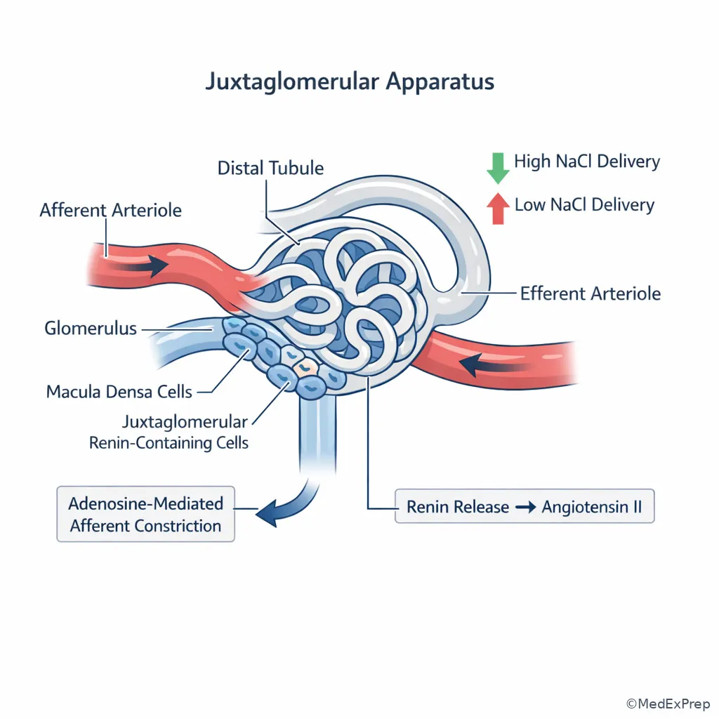
Renal autoregulation at the macula densa
Concept flow: Predict the drug-hemodynamic effect
Check baseline perfusion dependence
Ask whether the patient already depends on prostaglandins or angiotensin II to maintain GFR.

Identify the drug target
NSAID blocks afferent rescue; ACEi/ARB removes efferent rescue; both together magnify risk.

Predict consequence
Expect creatinine rise if compensatory autoregulation is disabled in a vulnerable hemodynamic state.

Key rule: A small creatinine rise after starting ACE inhibitor in proteinuric CKD can be acceptable, but a larger rise in bilateral renal artery stenosis, severe hypovolemia, or decompensated heart failure should trigger reassessment of renal perfusion and medications.
High-Yield Connection: This concept overlaps with Resistant And Secondary Hypertension and Prerenal And Hemodynamic AKI. Renovascular disease questions often hinge on the same efferent-support logic tested here.
Clinician Action: Diagnose drug-related hemodynamic AKI by timing, medication exposure, low effective arterial blood volume, and bland urine sediment. Order medication reconciliation, orthostatic or hemodynamic assessment, urinalysis, and selective renovascular evaluation if clinically suggested. Treat by correcting perfusion, holding the offending drug when appropriate, and restarting thoughtfully once stable. Refer/Escalate if oliguria, refractory hyperkalemia, or severe renal artery stenosis is suspected.

B Worked example

A 76-year-old woman with long-standing hypertension, peripheral vascular disease, heart failure with reduced ejection fraction, and stage 3 CKD is started on lisinopril for albuminuric kidney disease. One week later, creatinine rises from 1.6 to 2.4 mg/dL and potassium is 5.3 mEq/L. She denies rash or fever. Urinalysis shows no blood and no pyuria. A renal bruit is noted, but the patient also reports poor oral intake after a dental procedure, making volume depletion a tempting alternative explanation.

Identify task: choose the most likely mechanism of the creatinine rise. Extract key facts: ACE inhibitor initiation, vascular disease, bruit, bland urine, mild hyperkalemia. Apply rule: efferent dilation after ACE inhibitor lowers glomerular capillary pressure; this effect is exaggerated when renal perfusion depends on angiotensin II, as in bilateral renal artery stenosis. Eliminate distractors: absence of pyuria/rash argues against AIN; no muddy brown casts are provided for ATI; pure dehydration is possible but does not explain the bruit clue as cleanly. Verify: the most likely mechanism is loss of angiotensin II-mediated efferent arteriolar constriction in renovascular disease.

C Exam trap

Exam trap: Calling any creatinine rise after ACE inhibitor “acute tubular necrosis from the drug.” WHY IT IS TEMPTING: the timing after drug initiation feels causal. SINGLE CLUE: bland urine sediment with renovascular clues points to hemodynamic fall in intraglomerular pressure, not tubular necrosis.

D Checkpoint

Question: In a patient with advanced cirrhosis and low effective arterial blood volume, which medication is most likely to cause an abrupt fall in GFR by preventing compensatory afferent vasodilation?

  1. Ibuprofen
  2. Lisinopril
  3. Spironolactone
  4. Amlodipine

Answer: A

  • A: Correct because NSAIDs inhibit prostaglandin synthesis, removing afferent vasodilatory support in low-perfusion states.
  • B: Tempting because ACE inhibitors can also lower GFR, but they do so primarily by efferent dilation rather than by blocking afferent prostaglandins.
  • C: Tempting in cirrhosis because it affects hemodynamics and potassium, but it is not the classic afferent constrictor mechanism tested here.
  • D: Tempting because it is a vasodilator, but it does not target the renal prostaglandin pathway responsible for this autoregulatory support.
If you missed this: Review 1.1.1.3 Autoregulation and vasoactive drugs — focus on NSAID effect on prostaglandin-dependent afferent vasodilation.

1.1.1.6 Measuring kidney function: true GFR, estimated GFR, and when creatinine misleads you

A Explanation

The decision conflict here is whether to trust the serum creatinine at face value. The single rule: creatinine reflects filtration and creatinine generation, distribution, and secretion. Therefore, creatinine is most reliable in steady state and least reliable when muscle mass is unusual or kidney function is changing rapidly.

Measured GFR is rarely needed on routine wards, but the physiology matters. Inulin clearance is ideal but impractical. Timed creatinine clearance is more available but imperfect because tubular secretion overestimates true GFR. Estimated GFR equations based on serum creatinine are extremely useful for outpatient CKD staging and medication adjustment, but not for abrupt AKI because creatinine lags behind the true fall in filtration. In a rapidly evolving injury, a “normal” creatinine may still coexist with a markedly reduced true GFR.

Cystatin C is useful when creatinine is unreliable, including low muscle mass, amputation, malnutrition, frailty, and sometimes cirrhosis. However, cystatin C is not perfect; inflammation, thyroid disease, and glucocorticoids can affect it. The exam usually rewards the broader principle rather than obscure exceptions. See Fig 5.

WHY THIS IS TESTED: many board stems ask for the best test to assess kidney function in cachexia, acute illness, obesity extremes, amputation, or cirrhosis.

Fig 5. Choosing the right filtration marker
Exam takeaway: Use creatinine-based eGFR in steady state, question it in unstable or low-muscle states, and escalate to alternative markers or measured approaches when the estimate is likely misleading.
flowchart TD
A[Need kidney function assessment] --> B{Steady state?}
B -->|Yes| C{Muscle mass typical?}
B -->|No| D[Do not rely on eGFR alone in AKI]
C -->|Yes| E[Creatinine-based eGFR usually acceptable]
C -->|No| F[Consider cystatin C or measured clearance]
D --> G[Use trend, urine data, and clinical context]
F --> H[Interpret with awareness of non-GFR influences]
Creatinine versus cystatin C comparison panel
Exam takeaway: Low muscle mass can falsely reassure with creatinine, making cystatin C or measured approaches more informative.
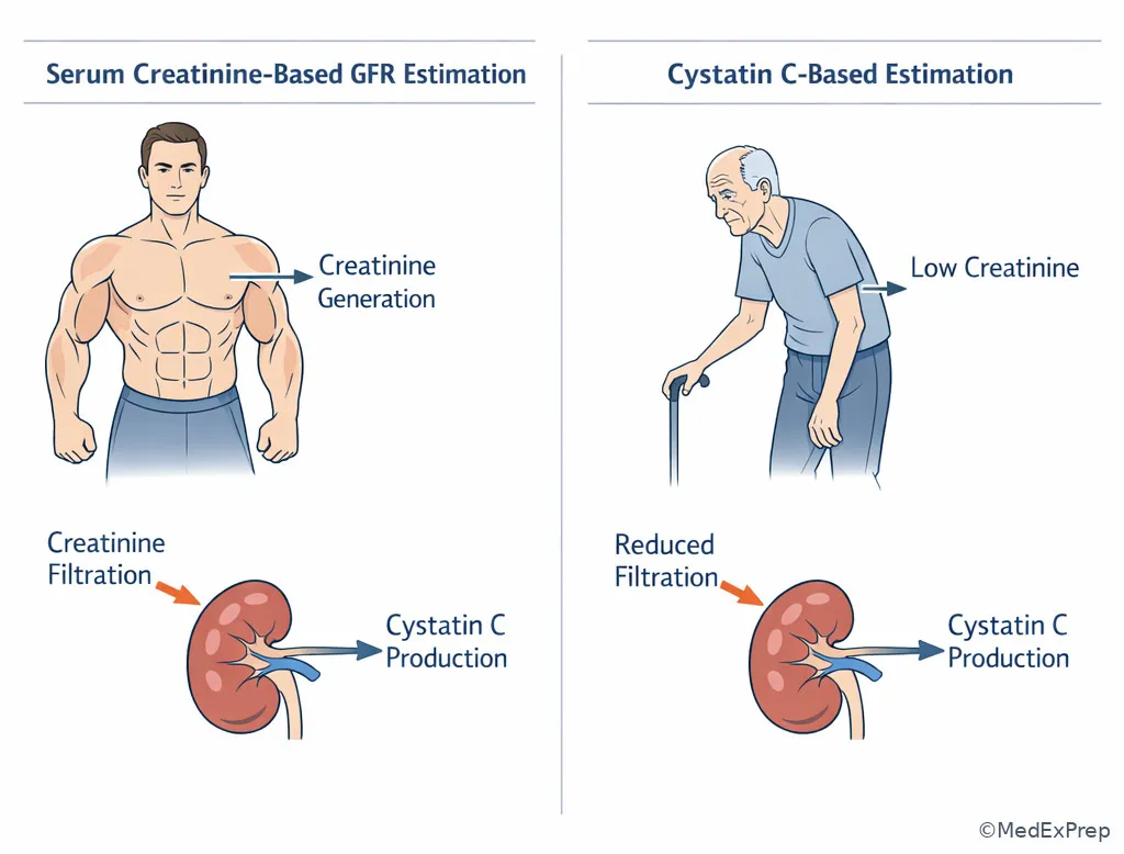
Creatinine versus cystatin C comparison panel
Concept flow: Can you trust this creatinine?
Check for steady state
Rapidly changing kidney function makes eGFR formulas unreliable.

Check creatinine generation
Low or high muscle mass can push creatinine away from true filtration status.

Choose alternative if needed
Use cystatin C or measured clearance when the estimate could mislead management.

Memory anchor: CREAT for creatinine pitfalls: Changing kidney function, Reduced muscle mass, Excess muscle breakdown, Amputation, Tubular secretion/drug effects.
Recall: In the upcoming topic Urine Concentration And Dilution, serum markers alone will not answer water-balance questions. Here, we build the habit of asking whether the filtration estimate itself is trustworthy before interpreting downstream urine data.
Clinician Action: Diagnose whether creatinine truly reflects filtration by checking for steady state and muscle-mass mismatch. Order serial creatinine, cystatin C when appropriate, medication review for secretion-interfering agents, and measured clearance only when it will change management. Treat the clinical kidney problem, not the number in isolation. Refer/Escalate if discordant markers create major uncertainty around dosing, biopsy decisions, or dialysis planning.

B Worked example

A 71-year-old woman with cirrhosis, severe sarcopenia, metastatic breast cancer, and chronic edema is referred for “normal kidney function” because her creatinine is 0.6 mg/dL. She is being considered for a renally cleared chemotherapy adjunct. Blood pressure is low-normal. Urinalysis is bland. A trainee is reassured by the low creatinine and wants standard dosing.

Identify task: determine whether creatinine-based eGFR is reliable. Extract key facts: severe sarcopenia, cirrhosis, malignancy, bland urinalysis, low creatinine. Apply rule: low creatinine generation can mask substantial reduction in true GFR. Eliminate distractors: bland urinalysis does not prove normal filtration; edema is not enough to classify renal function. Verify: the most appropriate next step is to use an alternative filtration assessment, often cystatin C-based estimation, before making dosing decisions.

C Exam trap

Exam trap: Equating “low creatinine” with “good kidney function.” WHY IT IS TEMPTING: creatinine is commonly taught as inverse to GFR. SINGLE CLUE: severe sarcopenia or cirrhosis breaks that assumption because creatinine production is reduced.

D Checkpoint

Question: Which setting most strongly limits the accuracy of creatinine-based eGFR for estimating true kidney function?

  1. Stable outpatient CKD with unchanged muscle mass over months
  2. Acute septic shock with rapidly rising creatinine over 12 hours
  3. Controlled hypertension with trace albuminuria
  4. Normal-weight adult with routine annual labs

Answer: B

  • A: Tempting because CKD is where GFR matters, but steady-state outpatient CKD is precisely where eGFR performs best.
  • B: Correct because creatinine lags behind abrupt changes in filtration, making formula-based eGFR unreliable in evolving AKI.
  • C: Tempting because albuminuria complicates CKD care, but it does not inherently invalidate creatinine-based eGFR.
  • D: Tempting because screening settings can hide mild disease, but the equation itself is generally more valid here than in unstable illness.
If you missed this: Review 1.1.1.4 Measuring kidney function — focus on the requirement for steady-state creatinine when using eGFR formulas.
Quick Recall:

  • The ideal measured GFR marker is ______.
  • True or False: Creatinine clearance slightly overestimates true GFR.
  • Name one setting where cystatin C may outperform creatinine.

1.1.1.7 Integration: hemodynamic patterns in real nephrology vignettes

A Explanation

The final decision conflict is which hemodynamic pattern best explains the entire vignette and therefore what the most appropriate next step is. The single rule is to integrate the volume state, medication exposures, urine sediment, and comorbid conditions before labeling the problem as intrinsic renal disease.

In effective arterial underfilling states such as heart failure and cirrhosis, total body fluid may be increased while the kidney senses low perfusion. This distinction is tested repeatedly. The kidney activates sympathetic tone, RAAS, and vasopressin; renal plasma flow falls and compensatory efferent constriction helps sustain filtration. Add NSAIDs or excessive diuresis and GFR falls. In bilateral renal artery stenosis, perfusion pressure distal to the stenosis is low, so angiotensin II support becomes essential; ACE inhibitor can therefore unmask a marked decline in GFR. In obstruction, the clue is rising Bowman space pressure rather than a vascular mechanism. In intrinsic glomerular or tubular disease, the sediment usually becomes more informative than the drug list alone.

The exam also likes “most important to monitor” questions. After starting RAAS blockade, monitor creatinine and potassium. After NSAID exposure in a vulnerable patient, monitor kidney function and volume status. In suspected hemodynamic AKI, the next step is often not biopsy but restoration of perfusion, drug review, and reassessment. See Fig 6.

WHY THIS IS TESTED: nephrology boards emphasize mechanism-to-management logic: know the physiologic reason first, then choose the safest next action.

Fig 6. High-yield hemodynamic differential for falling GFR
Exam takeaway: Volume state, vasoactive medications, urine sediment, and obstruction clues separate hemodynamic AKI from intrinsic renal disease.
flowchart TD
A[Falling GFR / rising creatinine] --> B{Urine sediment active?}
B -->|Yes| C[Think intrinsic renal disease]
B -->|No| D{Hemodynamic clue present?}
D -->|NSAID + low EABV| E[Afferent constriction]
D -->|ACEi/ARB + renovascular state| F[Efferent dilation]
D -->|HF/cirrhosis/hypovolemia| G[Reduced renal perfusion with compensation]
D -->|Hydronephrosis / retention| H[Obstruction]
E --> I[Hold trigger, restore perfusion]
F --> I
G --> I
H --> J[Relieve obstruction urgently]
Hemodynamic AKI comparison panel
Exam takeaway: Side-by-side visualization of hypovolemia, heart failure, cirrhosis, and bilateral renal artery stenosis helps identify which compensatory pathway the kidney is relying on.
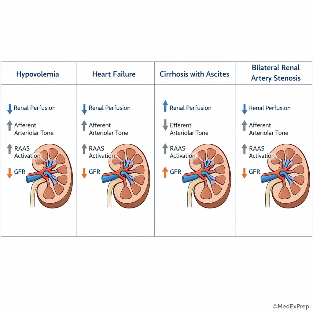
Hemodynamic AKI comparison panel
Hydronephrosis and back-pressure effect on filtration
Exam takeaway: Obstruction lowers GFR by increasing Bowman space pressure rather than by primary afferent or efferent arteriolar tone changes.
Hydronephrosis and back-pressure effect on filtration
Concept flow: Most appropriate first-pass interpretation of rising creatinine
Review context
Look for low effective arterial volume, offending drugs, and obstruction clues.

Check sediment
Bland urine favors hemodynamic or obstructive disease; active sediment pushes toward intrinsic pathology.

Act on mechanism
Restore perfusion, remove triggers, or relieve obstruction before chasing rare diagnoses.

Clinician Action: Diagnose hemodynamic versus intrinsic versus obstructive causes of falling GFR using the bedside triad of volume state, medication exposure, and urine sediment. Order urinalysis, urine microscopy, medication review, bladder scan or kidney ultrasound when obstruction is possible, and targeted labs for intrinsic disease only when supported. Treat by removing the offending hemodynamic hit, optimizing perfusion, and addressing obstruction rapidly. Refer/Escalate for severe AKI, uncertain diagnosis, rapidly active sediment, or dialysis indications.

B Worked example

A 64-year-old man with ischemic cardiomyopathy, BPH, and CKD stage 3 presents with dyspnea and edema. He receives aggressive diuresis and several doses of ketorolac for back pain. Creatinine rises from 1.8 to 2.7 mg/dL. He has mild suprapubic fullness but passes some urine. Urinalysis shows hyaline casts only. Ultrasound later shows mild bilateral hydronephrosis, while exam also suggests persistent venous congestion. The tempting wrong answer is “ATN from overdiuresis” because the creatinine rose after hospitalization.

Identify task: select the most appropriate first-pass mechanism and next step. Extract key facts: CHF with low effective arterial blood volume, NSAID exposure, BPH, bland sediment, possible obstruction. Apply rule: bland sediment plus multiple hemodynamic/obstructive hits argues against primary intrinsic tubular injury. Eliminate distractors: hyaline casts are nonspecific; absence of muddy brown casts makes ATI less compelling. Verify: the best next step is to evaluate and relieve obstruction while stopping NSAIDs and reassessing hemodynamics, because obstruction and hemodynamic compromise are both more likely than intrinsic necrosis.

C Exam trap

Exam trap: Choosing biopsy or serologic workup first in any unexplained creatinine rise. WHY IT IS TEMPTING: nephrology exams often teach broad differentials, and candidates can over-advance to rare diagnoses. SINGLE CLUE: a bland sediment plus obvious hemodynamic or obstructive clues means the safer first move is mechanism-based bedside evaluation.

D Checkpoint

Question: A patient with decompensated heart failure has edema, low urine sodium, bland urine sediment, and rising creatinine after ibuprofen exposure. What is the most likely immediate mechanism of the GFR decline?

  1. Loss of prostaglandin-mediated afferent vasodilation
  2. Immune complex deposition in the mesangium
  3. Myoglobin-induced tubular obstruction
  4. Decreased creatinine production from sarcopenia

Answer: A

  • A: Correct because low effective arterial blood volume makes the kidney prostaglandin dependent, and NSAID exposure removes afferent vasodilatory support.
  • B: Tempting because kidney dysfunction can reflect glomerular disease, but the bland sediment and drug/volume clues point away from inflammatory pathology.
  • C: Tempting because tubular obstruction is a renal mechanism, but there is no rhabdomyolysis context or pigmented casts.
  • D: Tempting because creatinine interpretation matters, but decreased production would falsely lower creatinine rather than explain its acute rise.
If you missed this: Review 1.1.1.5 Integration: hemodynamic patterns in real nephrology vignettes — focus on bland sediment plus NSAID exposure in low effective arterial blood volume.
TRAPS

Exam Traps & Differentiators

Common traps & distractors:

  • Most common wrong answer: intrinsic renal disease when the real problem is hemodynamic. Why: creatinine rose and the stem feels “renal.” Fix: first ask whether the urine sediment is bland and whether a drug/volume explanation is sitting in front of you.
  • Looks similar but isn't: cirrhosis and heart failure can present with edema yet still produce low effective arterial blood volume; the kidney responds as if underperfused.
  • False reassurance trap: low creatinine in sarcopenia or cirrhosis. The exam wants you to distrust a deceptively normal creatinine when muscle mass is low.
  • Drug-mechanism swap: candidates reverse NSAID and ACE inhibitor effects. NSAIDs hit the afferent rescue pathway; ACE inhibitors remove efferent rescue.
Look-alike scenario Why it tempts you Decisive clue Think
CHF with rising creatinine after NSAID Hospitalized patient with AKI could be ATI Bland sediment, low effective arterial volume, NSAID timing Afferent constriction/hemodynamic AKI
Renal bruit and creatinine rise after ACE inhibitor Medication-related AKI may sound nonspecific Vascular disease plus marked creatinine increase after RAAS blockade Loss of efferent support in renovascular disease
Edema with low urine sodium in cirrhosis Edema suggests volume overload only Kidney senses low effective arterial blood volume despite excess total body fluid Perfusion-dependent RAAS activation
Low serum creatinine in cachexia Low creatinine feels “good” Severe muscle loss reduces creatinine generation eGFR may overestimate true kidney function
AKI with hydronephrosis Could still have medications on board Back-pressure clues or bladder outlet symptoms Obstructive fall in GFR
If the stem says... Think...
“Started ibuprofen during GI illness” Loss of prostaglandin-mediated afferent vasodilation
“Creatinine rose after ACE inhibitor and there is a renal bruit” Bilateral renal artery stenosis or severe renovascular dependence on angiotensin II
“Massive edema but kidney behaves as if underfilled” Low effective arterial blood volume, not true adequate perfusion
“Bland sediment and clear hemodynamic insult” Choose hemodynamic AKI before intrinsic pathology
“Sarcopenic patient with very low creatinine” Creatinine may overestimate GFR; consider alternative markers
“Hydronephrosis or urinary retention” Increased Bowman space pressure lowering GFR

Related trap from upcoming topics: when you later study Fractional Excretion Reasoning, do not overread FeNa in patients on diuretics or mixed hemodynamic-intrinsic states. The hemodynamic logic from this lesson comes first; urine indices refine it.

Review connection: The traps above test renal hemodynamic interpretation. If any felt unfamiliar, revisit 1.1.1.3 Autoregulation and vasoactive drugs and 1.1.1.5 Integration: hemodynamic patterns in real nephrology vignettes before attempting the Self-check quiz.
ALGO

Algorithm / Approach

Concept flow: Approach to rising creatinine through GFR and renal blood flow
Start
Confirm whether kidney function is changing acutely or the patient is in steady state.

Decision
If urine sediment is bland, prioritize hemodynamic and obstructive causes; otherwise evaluate intrinsic disease.

Medication check
Identify NSAIDs, ACE inhibitors, ARBs, diuretics, and other agents that alter afferent/efferent tone or perfusion.

Interpret mechanism
Low effective arterial blood volume + NSAID suggests afferent problem; renovascular dependence + ACEi/ARB suggests efferent problem; retention/hydronephrosis suggests back-pressure.

Next step
Restore perfusion, remove the trigger, relieve obstruction, and only then escalate to intrinsic workup if the pattern does not fit or the sediment is active.

Clinical approach to a creatinine rise after medication exposure
Exam takeaway: A structured medication-volume-sediment review often answers the stem before advanced testing is needed.
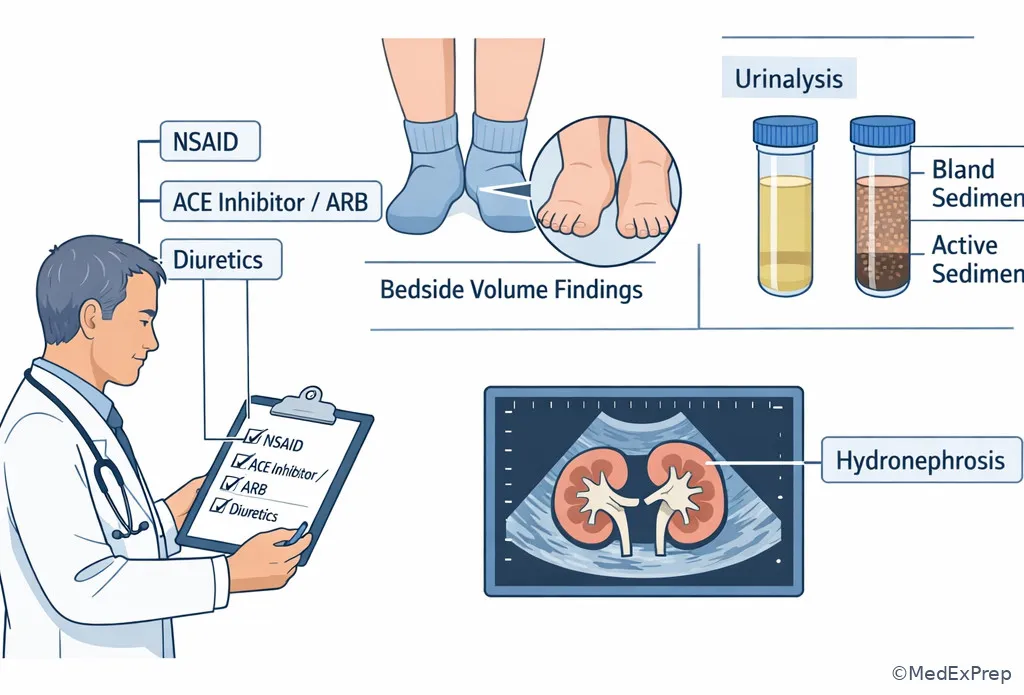
Clinical approach to a creatinine rise after medication exposure
RAPID

Rapid Review

  • Afferent constriction → RBF down, GFR down
  • Afferent dilation → RBF up, GFR up
  • Efferent constriction → RPF down, FF up
  • Efferent dilation → intraglomerular pressure down, GFR down
  • Obstruction → Bowman space pressure up
  • PAH clearance → approximates RPF, not GFR
  • Inulin clearance → ideal measured GFR marker
  • Creatinine clearance → slight GFR overestimate from secretion
  • AKI in non–steady state → eGFR formula unreliable
  • Sarcopenia/cirrhosis → creatinine may overestimate kidney function
  • NSAID-associated AKI → prostaglandin-dependent afferent support lost
  • ACEi/ARB creatinine rise → efferent support withdrawn; watch for renovascular disease
  • CHF/cirrhosis edema → low effective arterial blood volume can still dominate renal behavior
  • Bland sediment → prioritize hemodynamic/obstructive causes before inflammatory intrinsic disease
Memory anchor: FLOW before FANCY — before choosing biopsy, serologies, or rare diagnoses, ask whether the stem is really about renal blood flow, afferent/efferent tone, or back-pressure.
QUIZ

Self-check quiz

1. A 45-year-old man undergoing physiology testing has a substance infused that is freely filtered and neither reabsorbed nor secreted. Which clearance most directly measures GFR?

  1. Inulin clearance
  2. PAH clearance
  3. Creatinine clearance
  4. Urea clearance

2. A 52-year-old woman with no kidney disease asks which vascular change most directly lowers GFR by reducing blood delivery into the glomerulus. Which is the best answer?

  1. Afferent arteriolar constriction
  2. Afferent arteriolar dilation
  3. Efferent arteriolar constriction
  4. Efferent arteriolar dilation

3. A 67-year-old man with HFrEF, diabetes, and osteoarthritis develops AKI after gastroenteritis. Medications are lisinopril, furosemide, and ibuprofen. Blood pressure is 90/56 mm Hg, urinalysis is bland, and creatinine rises from 1.1 to 2.0 mg/dL. What is the most appropriate physiologic explanation?

  1. Immune-mediated interstitial inflammation from ibuprofen
  2. Combined loss of afferent and efferent support of glomerular pressure in a low-perfusion state
  3. Postinfectious glomerulonephritis causing abrupt reduction in filtration coefficient
  4. Rhabdomyolysis causing pigmented cast nephropathy

4. A 74-year-old woman with peripheral vascular disease, resistant hypertension, and CKD stage 3 is started on losartan. One week later, creatinine rises by 35% and potassium is 5.4 mEq/L. She has a renal bruit and bland urine sediment. What is the most appropriate interpretation?

  1. Expected acute interstitial nephritis from ARB exposure
  2. Loss of angiotensin II-mediated efferent arteriolar constriction in renovascular disease
  3. Mesangial immune-complex disease unmasked by blood pressure control
  4. PAH secretion failure causing pseudoelevation of creatinine

5. A 70-year-old woman with cirrhosis, sarcopenia, and chronic edema has a creatinine of 0.5 mg/dL before planned dosing of a renally cleared drug. Blood pressure is 98/60 mm Hg, urinalysis is bland, and there is marked muscle wasting. What is the most appropriate next step for assessing kidney function?

  1. Assume normal GFR because creatinine is low
  2. Use creatinine-based eGFR alone because cirrhosis does not affect creatinine generation
  3. Consider cystatin C-based estimation because creatinine may overestimate GFR in severe sarcopenia
  4. Delay all assessment until a kidney biopsy is performed

6. A 63-year-old man with decompensated cirrhosis, tense ascites, and CKD stage 2 undergoes large-volume paracentesis without adequate albumin replacement. Creatinine rises over 24 hours, urine sediment is bland, and urine sodium is low. Which renal hemodynamic pattern is most likely present early in this course?

  1. Increased renal plasma flow and decreased filtration fraction
  2. Decreased renal plasma flow with preserved or relatively maintained GFR from efferent constriction
  3. Increased Bowman space pressure from occult obstruction
  4. Normal renal plasma flow with decreased GFR from isolated fall in filtration coefficient

7. A 68-year-old woman with CHF, BPH, CKD stage 3, and chronic back pain presents with rising creatinine after aggressive diuresis and ketorolac exposure. She has mild suprapubic fullness, still makes urine, and urinalysis shows only hyaline casts. Kidney ultrasound reveals new bilateral hydronephrosis. What is the most appropriate next step?

  1. Start high-dose corticosteroids for presumed interstitial nephritis
  2. Order urgent measures to relieve obstruction while stopping hemodynamically harmful medications
  3. Continue NSAIDs because the bland urine excludes hemodynamic injury
  4. Proceed directly to kidney biopsy because hydronephrosis does not affect GFR

8. A 60-year-old man with bilateral renal artery stenosis, diabetes, and albuminuric CKD is started on an ACE inhibitor. His creatinine rises substantially, but urine sediment remains bland and there is no hypotension. Which single physiologic detail best distinguishes this from simple volume depletion?

  1. The ACE inhibitor causes afferent constriction instead of efferent dilation
  2. Renal perfusion beyond the stenoses depends on angiotensin II to maintain intraglomerular pressure
  3. Creatinine rises only in intrinsic tubular necrosis, not hemodynamic states
  4. Filtration fraction must decrease in all RAAS-activated states
ANSWERS

Answer key

1. Correct answer: A — Inulin clearance.

  • A: Tempting because it is the classic textbook answer, and it is correct here because inulin is freely filtered without reabsorption or secretion, making its clearance equal to GFR. Single clue: “freely filtered and neither reabsorbed nor secreted.” Source: Brenner and Rector’s The Kidney; Harrison’s Principles of Internal Medicine.
  • B: Tempting because PAH is another famous renal physiology marker, but it approximates effective renal plasma flow due to filtration plus secretion. Single clue: the stem asks for direct measurement of GFR, not plasma flow. Source: Brenner and Rector; Costanzo Physiology.
  • C: Tempting because creatinine is used clinically to estimate GFR, but secretion makes creatinine clearance a slight overestimate rather than the ideal direct measure. Single clue: “directly measures” excludes the imperfect surrogate. Source: KDIGO CKD guideline; Harrison’s.
  • D: Tempting because urea is filtered and familiar in nephrology, but variable tubular reabsorption makes it unreliable for direct GFR measurement. Single clue: reabsorption violates the ideal marker rule. Source: Costanzo Physiology; Brenner and Rector.
If you missed this: Review 1.1.1.2 Renal blood flow, renal plasma flow, and filtration fraction — focus on which clearance marker corresponds to which renal physiologic variable.

2. Correct answer: A — Afferent arteriolar constriction.

  • A: Tempting because it is the simplest inflow mechanism, and it is correct because narrowing the vessel entering the glomerulus reduces renal blood flow and glomerular capillary hydrostatic pressure. Single clue: “reducing blood delivery into the glomerulus.” Source: Costanzo Physiology; Harrison’s.
  • B: Tempting because arteriolar changes often alter GFR, but afferent dilation increases inflow and usually raises GFR. Single clue: dilation has the opposite direction of effect. Source: Brenner and Rector.
  • C: Tempting because efferent constriction certainly changes filtration dynamics, but mild-to-moderate efferent constriction tends to preserve or raise glomerular pressure rather than directly reducing inflow. Single clue: the stem emphasizes delivery into, not pressure distal to, the glomerulus. Source: Harrison’s; Costanzo.
  • D: Tempting because efferent dilation lowers GFR, but it does so by lowering intraglomerular pressure, not by reducing inflow into the tuft. Single clue: vessel leaving the glomerulus is not the inflow resistor. Source: Brenner and Rector.
If you missed this: Review 1.1.1.1 Foundational determinants: what sets GFR at the glomerulus? — focus on afferent versus efferent roles in inflow and intraglomerular pressure.

3. Correct answer: B — Combined loss of afferent and efferent support of glomerular pressure in a low-perfusion state.

  • A: Tempting because ibuprofen can cause AIN, but the absence of pyuria, rash, fever, and active sediment makes immune interstitial injury less likely in this specific case. Single clue: bland urinalysis with hypotension and combined NSAID/ACEi exposure. Source: Harrison’s; UpToDate review of NSAID nephrotoxicity.
  • B: Correct because hypovolemia lowers renal perfusion, ibuprofen blocks prostaglandin-dependent afferent vasodilation, and lisinopril removes angiotensin II-mediated efferent constriction; together these sharply reduce glomerular filtration pressure. Single clue: gastroenteritis plus ibuprofen plus lisinopril with bland sediment. Source: KDIGO AKI guideline; Brenner and Rector.
  • C: Tempting because infection precedes some glomerulonephritides, but there is no nephritic sediment, edema pattern, or complement clue here. Single clue: bland sediment argues against inflammatory glomerular injury. Source: Harrison’s.
  • D: Tempting because AKI can result from pigmented casts, but the stem offers no muscle injury, dark urine, or hyperCKemia clue. Single clue: the medication-volume story fully explains the presentation. Source: KDIGO AKI guideline; Harrison’s.
If you missed this: Review 1.1.1.3 Autoregulation and vasoactive drugs — focus on NSAID effect on afferent tone and ACE inhibitor effect on efferent tone in low-perfusion states.

4. Correct answer: B — Loss of angiotensin II-mediated efferent arteriolar constriction in renovascular disease.

  • A: Tempting because any new medication can cause drug-related AKI, but ARBs are not classic triggers of AIN, and bland urine with vascular clues points elsewhere. Single clue: renal bruit and resistant hypertension. Source: Harrison’s; AHA/ACC hypertension guidance.
  • B: Correct because in bilateral renovascular disease, downstream perfusion pressure is low and angiotensin II-dependent efferent constriction helps sustain GFR; blocking this causes a marked creatinine rise. Single clue: rise after losartan in a patient with vascular disease and renal bruit. Source: Brenner and Rector; Harrison’s.
  • C: Tempting because CKD can coexist with glomerular disease, but there is no hematuria, proteinuria pattern, or active sediment supporting immune-complex pathology. Single clue: the timing after ARB initiation fits a hemodynamic explanation far better. Source: Harrison’s.
  • D: Tempting because secretion pathways matter for renal markers, but PAH is a physiology test substance and not the mechanism of this clinical creatinine change. Single clue: real-world ARB exposure with renovascular clues. Source: Costanzo Physiology.
If you missed this: Review 1.1.1.3 Autoregulation and vasoactive drugs — focus on why renovascular states depend on angiotensin II-mediated efferent support.

5. Correct answer: C — Consider cystatin C-based estimation because creatinine may overestimate GFR in severe sarcopenia.

  • A: Tempting because a low creatinine superficially suggests preserved filtration, but severe sarcopenia lowers creatinine production and can mask major kidney dysfunction. Single clue: marked muscle wasting. Source: KDIGO CKD guideline; Harrison’s.
  • B: Tempting because creatinine-based eGFR is commonly used, but cirrhosis and sarcopenia both reduce reliability of creatinine-based estimation. Single clue: the stem intentionally pairs cirrhosis with muscle loss. Source: KDIGO; Brenner and Rector.
  • C: Correct because alternative filtration markers such as cystatin C can be more informative when creatinine generation is abnormally low. Single clue: low creatinine in the setting of profound sarcopenia. Source: KDIGO CKD guideline; Harrison’s.
  • D: Tempting because definitive evaluation sounds thorough, but biopsy assesses kidney pathology, not first-line estimation of filtration in this context. Single clue: the question asks kidney function assessment for drug dosing, not tissue diagnosis. Source: KDIGO; Harrison’s.
If you missed this: Review 1.1.1.4 Measuring kidney function — focus on why low muscle mass makes serum creatinine falsely reassuring.

6. Correct answer: B — Decreased renal plasma flow with preserved or relatively maintained GFR from efferent constriction.

  • A: Tempting because hyperdynamic cirrhosis can confuse total circulatory concepts, but the kidney in low effective arterial volume does not experience increased renal plasma flow. Single clue: low urine sodium and post-paracentesis underfilling imply renal vasoconstrictive compensation. Source: Harrison’s; UpToDate review of hepatorenal physiology.
  • B: Correct because RAAS activation lowers renal plasma flow while angiotensin II-mediated efferent constriction initially helps maintain filtration pressure, often increasing filtration fraction. Single clue: early bland AKI after inadequate albumin replacement in cirrhosis. Source: Brenner and Rector; Harrison’s.
  • C: Tempting because obstruction lowers GFR with bland sediment, but there is no retention or hydronephrosis clue; the timing after paracentesis points to circulatory underfilling. Single clue: procedure-related effective volume loss. Source: Harrison’s.
  • D: Tempting because filtration coefficient can fall in intrinsic disease, but the vignette is hemodynamic rather than glomerular inflammatory. Single clue: low urine sodium and cirrhotic underfilling. Source: KDIGO AKI guideline; Harrison’s.
If you missed this: Review 1.1.1.2 Renal blood flow, renal plasma flow, and filtration fraction — focus on the rise in filtration fraction during efferent-constrictive compensation.

7. Correct answer: B — Order urgent measures to relieve obstruction while stopping hemodynamically harmful medications.

  • A: Tempting because NSAIDs can cause AIN, but hydronephrosis and outlet symptoms supply a more direct and urgent explanation for falling GFR. Single clue: new bilateral hydronephrosis. Source: KDIGO AKI guideline; Harrison’s.
  • B: Correct because obstructive AKI can coexist with hemodynamic injury, and urgent relief of obstruction plus removal of NSAIDs addresses the highest-yield reversible causes first. Single clue: bilateral hydronephrosis in a patient with BPH and rising creatinine. Source: KDIGO; Brenner and Rector.
  • C: Tempting because bland urine does suggest hemodynamic rather than intrinsic injury, but it does not make NSAIDs safe; they remain harmful in this low-perfusion state. Single clue: ketorolac exposure in CHF and AKI. Source: Harrison’s.
  • D: Tempting because biopsy can clarify AKI, but obstruction is a known, reversible cause and hydronephrosis absolutely can lower GFR by increasing Bowman space pressure. Single clue: structural back-pressure is already demonstrated. Source: Harrison’s; Brenner and Rector.
If you missed this: Review 1.1.1.5 Integration: hemodynamic patterns in real nephrology vignettes — focus on why bland sediment plus hydronephrosis should redirect you to obstruction before intrinsic workup.

8. Correct answer: B — Renal perfusion beyond the stenoses depends on angiotensin II to maintain intraglomerular pressure.

  • A: Tempting because students mix up arteriolar drug effects, but ACE inhibitors cause efferent dilation, not afferent constriction. Single clue: RAAS blockade acts on angiotensin II-mediated efferent tone. Source: Costanzo Physiology; Harrison’s.
  • B: Correct because the stenotic kidneys rely on angiotensin II-mediated efferent constriction to preserve glomerular capillary hydrostatic pressure despite reduced inflow pressure. Single clue: bilateral renal artery stenosis with bland sediment after ACE inhibitor initiation. Source: Brenner and Rector; AHA/ACC guidance.
  • C: Tempting because large creatinine rises feel like structural injury, but hemodynamic states can produce substantial increases without tubular necrosis. Single clue: bland sediment argues against intrinsic tubular injury. Source: KDIGO AKI guideline.
  • D: Tempting because filtration fraction is a hemodynamic concept linked to RAAS, but it does not necessarily decrease in all RAAS-activated states and often rises with efferent constriction. Single clue: the discriminating issue is dependence on angiotensin II, not a universal FF direction. Source: Costanzo; Harrison’s.
If you missed this: Review 1.1.1.3 Autoregulation and vasoactive drugs — focus on angiotensin II-dependent efferent support in bilateral renal artery stenosis.

References

  • Kidney Disease: Improving Global Outcomes (KDIGO). “KDIGO 2024 Clinical Practice Guideline for the Evaluation and Management of Chronic Kidney Disease.”
  • Kidney Disease: Improving Global Outcomes (KDIGO). “Clinical Practice Guideline for Acute Kidney Injury.”
  • Taal, Maarten W., et al., editors. Brenner and Rector’s The Kidney. 11th ed., Elsevier.
  • Jameson, J. Larry, et al., editors. Harrison’s Principles of Internal Medicine. 21st ed., McGraw-Hill.
  • Costanzo, Linda S. Physiology. 7th ed., Elsevier.
  • Whelton, Paul K., et al. “2017 ACC/AHA Guideline for the Prevention, Detection, Evaluation, and Management of High Blood Pressure in Adults.” Journal of the American College of Cardiology.

Last reviewed: March 15, 2026

Lesson 2

Tubular Transport Mechanisms

Tubular Transport Mechanisms

Course: ABIM Nephrology Prep Course | Chapter: Renal Physiology | Section: Glomerular And Tubular Physiology | Level: Foundational

Glomerular filtration creates tubular fluid; selective transport determines what ultimately appears in urine. For board examinations, recognizing where a solute is handled is often the shortest path from an electrolyte pattern to a mechanism, medication effect, or inherited transport defect.

What you'll learn

  • PHYSIOLOGY_PATHOLOGY_DIAGNOSTICS: Localize solute transport -> Distinguish proximal, thick ascending limb, distal convoluted tubule, and collecting duct functions.
  • PHYSIOLOGY_PATHOLOGY_DIAGNOSTICS: Explain epithelial transport -> Relate major transporters, electrochemical gradients, and hormonal regulation to urinary solute handling.
  • FLUID_ELECTROLYTE_ACID_BASE: Predict electrolyte consequences -> Anticipate potassium, calcium, magnesium, phosphate, bicarbonate, and water responses to segment-specific disturbances.
  • PHYSIOLOGY_PATHOLOGY_DIAGNOSTICS: Interpret pharmacologic inhibition -> Match diuretic classes to transport targets and characteristic adverse electrolyte patterns.
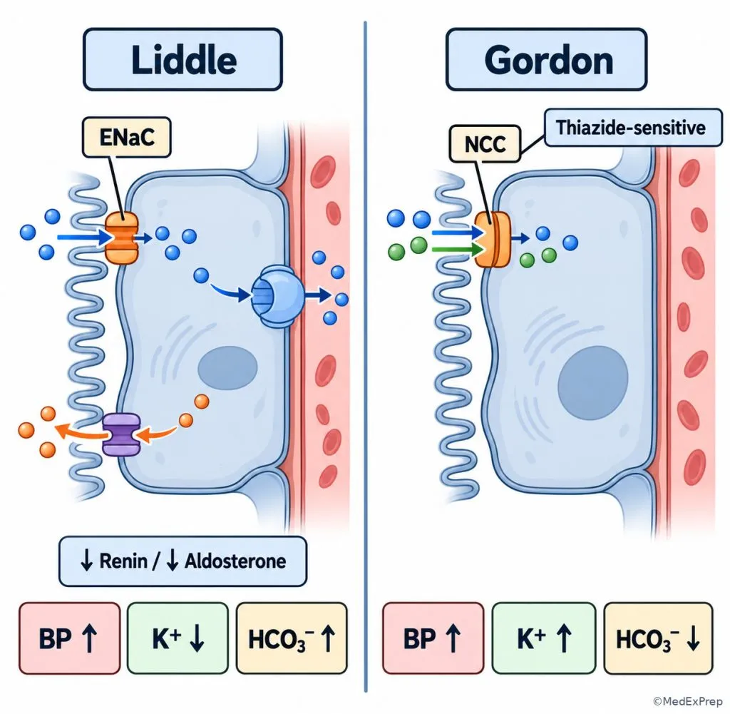
  • FLUID_ELECTROLYTE_ACID_BASE: Recognize transport disorders -> Differentiate proximal tubular dysfunction, Bartter-like, Gitelman-like, Liddle, and Gordon phenotypes.

Blueprint mapping: PHYSIOLOGY_PATHOLOGY_DIAGNOSTICS -> Renal physiology and diagnostics -> Tubular transport; FLUID_ELECTROLYTE_ACID_BASE -> Segmental electrolyte and water handling. Exact ABIM weighting for this subskill is not supplied.

Estimated study time: 50-65 minutes. Prerequisite: GFR And Renal Blood Flow. Key outputs: Choose proximal versus distal dysfunction, loop versus thiazide effects, aldosterone versus vasopressin regulation, and inherited versus acquired transport phenotypes.

Why it matters

  • Electrolyte abnormalities reflect a combination of filtered load, segmental reabsorption, secretion, hormone signaling, and delivery to downstream nephron segments.
  • The same serum potassium abnormality can arise from a diuretic, a hormone disorder, reduced distal sodium delivery, or a transport-channel defect; distinguishing them changes management.
  • Calcium and magnesium handling provides important discriminators when a patient has hypokalemic metabolic alkalosis or is receiving diuretic therapy.
  • Many inherited tubulopathies reproduce the physiologic effect of inhibiting a specific transporter, making medication patterns useful diagnostic analogies.

Key Terms & Must-Know Facts

Transcellular transport
Movement across apical and basolateral cell membranes through channels, carriers, or pumps.
Paracellular transport
Movement between epithelial cells through pathways influenced by permeability and electrochemical gradients.
Na+/K+-ATPase
Basolateral pump that maintains low intracellular sodium and supports secondary active sodium-coupled transport.
NHE3
Proximal apical sodium-hydrogen exchanger supporting sodium reabsorption and bicarbonate reclamation.
SGLT2
Proximal sodium-glucose cotransporter targeted by SGLT2 inhibitors.
NKCC2
Apical sodium-potassium-two-chloride cotransporter of the thick ascending limb; loop-diuretic target.
ROMK
Renal outer medullary potassium channel involved in potassium recycling in the thick ascending limb and potassium transport in the distal nephron.
NCC
Sodium-chloride cotransporter in the early distal convoluted tubule; thiazide target.
ENaC
Epithelial sodium channel of the aldosterone-sensitive distal nephron; amiloride-sensitive sodium entry pathway.
AQP2
Apical aquaporin recruited in collecting duct principal cells in response to vasopressin.
TRPV5
Apical calcium-entry channel participating in regulated distal calcium reabsorption.
TRPM6
Magnesium-entry channel important in distal convoluted tubule magnesium conservation.
Fanconi syndrome
Generalized proximal tubular reabsorptive dysfunction involving multiple filtered solutes.
Lumen-positive voltage
Electrical driving force in the thick ascending limb that promotes paracellular reabsorption of divalent cations.

Must-know facts

  • The proximal tubule reabsorbs most filtered sodium and water and nearly all filtered glucose under usual physiologic conditions.
  • Proximal bicarbonate reclamation depends substantially on sodium-hydrogen exchange and carbonic anhydrase activity.
  • The thick ascending limb reabsorbs salt while remaining relatively impermeable to water.
  • Loop inhibition promotes urinary calcium and magnesium losses; thiazide inhibition usually reduces urinary calcium excretion.
  • Distal potassium secretion depends on aldosterone action, distal sodium delivery, tubular flow, and potassium-channel function.
  • Vasopressin mainly regulates collecting duct water permeability, whereas aldosterone regulates sodium reabsorption and potassium secretion.
  • Generalized proximal transport failure produces multiple solute losses rather than isolated glucosuria.
  • Bartter-like and Gitelman-like disorders are usually salt-wasting, hypokalemic alkalosis phenotypes; Liddle and Gordon syndromes characteristically involve hypertension.
Table 1. Nephron segment transport reference
Segment Dominant transport mechanisms Water and electrolyte handling Board discriminator
Proximal tubule NHE3, SGLT transport, sodium-phosphate cotransport, carbonic anhydrase-dependent bicarbonate reclamation Bulk sodium, water, bicarbonate, glucose, phosphate and other solute reclamation Multiple urinary solute losses indicate generalized proximal dysfunction.
Thick ascending limb NKCC2 and ROMK NaCl reclamation without accompanying water; lumen-positive driving force for calcium and magnesium reabsorption Loop inhibition increases urinary calcium and magnesium loss.
Early distal convoluted tubule NCC, TRPV5-related calcium entry, TRPM6-related magnesium entry NaCl transport; regulated calcium and magnesium conservation Thiazide exposure or Gitelman phenotype favors reduced urinary calcium.
Connecting tubule and collecting duct ENaC, potassium channels, aldosterone-sensitive pathways, intercalated-cell acid-base transport, AQP2 Fine control of sodium, potassium, acid-base balance, and hormone-dependent water recovery Potassium and blood pressure patterns help distinguish hormone and channel disorders.

Core content

1. Proximal tubule: bulk recovery versus generalized solute wasting

A. Explanation

WHY THIS IS TESTED: A patient with glucosuria, low serum phosphate, or metabolic acidosis may have uncontrolled hyperglycemia, an isolated transporter effect, or generalized proximal tubule injury. The examination asks you to distinguish a single solute abnormality from coordinated loss of several substances that should normally be reclaimed.

The basolateral Na+/K+-ATPase generates the sodium gradient that supports proximal uptake through multiple apical transport mechanisms. SGLT transporters couple glucose uptake to sodium entry. NHE3 participates in sodium reabsorption and hydrogen secretion, which facilitates filtered bicarbonate reclamation through reactions involving carbonic anhydrase. Sodium-phosphate cotransporters reclaim phosphate; parathyroid hormone and FGF23 reduce proximal phosphate reabsorption. Water follows solute across a highly water-permeable epithelium, producing approximately isoosmotic bulk reabsorption.

Because the proximal tubule handles multiple filtered substrates, widespread dysfunction may produce euglycemic glucosuria, phosphaturia, bicarbonate wasting, aminoaciduria, and uricosuria. Not every component must be demonstrated in an individual patient. An isolated SGLT2 inhibitor effect produces glucosuria but does not by itself establish Fanconi syndrome. Likewise, proximal renal tubular acidosis describes impaired bicarbonate reclamation and does not automatically establish generalized proximal transport failure.

Decision rule: Choose a generalized proximal disorder when multiple unrelated filtered solutes are lost together; reject that diagnosis when only one substrate is affected and a selective transporter explanation is sufficient.

See Fig 1 for the nephron-wide localization map and Fig 2 for the cellular basis of proximal reclamation.

Step 1 - Identify urinary loss. Determine whether the abnormality involves glucose, bicarbonate, phosphate, or several solutes.
Step 2 - Check filtered-load explanations. Review serum glucose, serum phosphate, acid-base status, and kidney function.
Step 3 - Review selective drug effects. Identify SGLT2 inhibitors, carbonic anhydrase inhibitors, and potentially proximal-toxic exposures.
Step 4 - Localize. Multiple unexplained losses support generalized proximal dysfunction; isolated findings favor targeted mechanisms.
Fig 1. Transporter map along the nephron
Exam takeaway: Segment localization links a urinary electrolyte pattern to a transporter or medication class.
flowchart LR
A[Proximal: NHE3 SGLT phosphate] --> B[TAL: NKCC2 ROMK]
B --> C[Early DCT: NCC]
C --> D[Connecting tubule: ENaC]
D --> E[Collecting duct: AQP2 and acid-base transport]
Fig 2. Proximal epithelial transport
Exam takeaway: The basolateral sodium gradient supports reclamation of several filtered solutes through distinct apical pathways.
{"prompt":"High-accuracy adult nephrology textbook illustration of one proximal tubule epithelial cell between tubular lumen and peritubular capillary. Clean white background, crisp cool-blue and gray palette, thin outlines, minimal shading. Label apical brush border, NHE3, SGLT2, sodium-phosphate cotransporter, basolateral Na+/K+-ATPase, carbonic anhydrase, glucose, bicarbonate, phosphate, sodium, and water movement. Use anatomically logical arrows separating luminal uptake from basolateral exit. No brands, photorealism, ornamental objects, or gradients.","caption":"Proximal epithelial transport","size":"1024x1024"}

B. Worked example

A 53-year-old woman with type 2 diabetes, hypertension, and chronic hepatitis B reports fatigue. She receives tenofovir and recently started an SGLT2 inhibitor. Serum glucose is 108 mg/dL, bicarbonate is 18 mEq/L, and phosphate is reduced. Urine contains glucose despite euglycemia, with evidence of inappropriate renal phosphate loss. Trace leukocyte esterase is present without urinary symptoms.

  1. Identify the task: Determine whether glucosuria is an isolated pharmacologic effect or part of broader tubular dysfunction.
  2. Extract key facts: Euglycemic glucosuria coexists with phosphate wasting and metabolic acidosis; a potentially proximal-toxic medication is present.
  3. Apply the rule: Simultaneous defects in reclamation of different proximal substrates favor a Fanconi-type pattern.
  4. Eliminate alternatives: SGLT2 inhibition explains glucose loss but does not adequately explain the entire phosphate and bicarbonate pattern; asymptomatic trace leukocyte esterase does not localize the dysfunction.
  5. Verify: Assess additional proximal losses, confirm the acid-base mechanism, review medication exposure, and address potentially reversible causes.

Most likely localization: Generalized proximal tubular dysfunction, with medication-associated injury among the important etiologies to evaluate.

C. Exam trap

Common wrong answer: Attribute every abnormality to the SGLT2 inhibitor. Why it is tempting: Drug-associated glucosuria during normal serum glucose is expected. Single eliminating clue: Concurrent inappropriate phosphate wasting demonstrates a second proximal transport abnormality that isolated SGLT2 inhibition does not explain.

D. Checkpoint MCQ

A patient has euglycemic glucosuria, phosphaturia, bicarbonate wasting, and increased urinary amino acids. Which site is primarily affected?

  1. A. Thick ascending limb
  2. B. Proximal tubule
  3. C. Early distal convoluted tubule
  4. D. Collecting duct principal cell

Correct answer: B.

  • A: Thick ascending limb dysfunction can waste salt and divalent cations, making electrolyte loss plausible, but it does not produce this combined glucose-amino acid-bicarbonate pattern.
  • B: The proximal tubule reclaims all listed substrates, so a multisubstrate wasting phenotype localizes here.
  • C: Early distal disease can affect sodium, magnesium, and calcium, but substantial glucose and amino acid reclamation occurs upstream.
  • D: Principal cells regulate sodium, potassium, and water, but they do not perform bulk glucose and phosphate reclamation.
If you missed this: Review Proximal tubule: bulk recovery versus generalized solute wasting - focus on multisubstrate urinary loss as the segment-localizing finding.

2. Thick ascending limb and distal convoluted tubule: interpreting calcium and magnesium

A. Explanation

WHY THIS IS TESTED: Both loop and thiazide-associated disturbances may cause hypokalemic metabolic alkalosis. A patient may also have magnesium depletion. The decisive comparison is frequently calcium handling, because inhibiting transport in these segments has different effects on urinary calcium excretion.

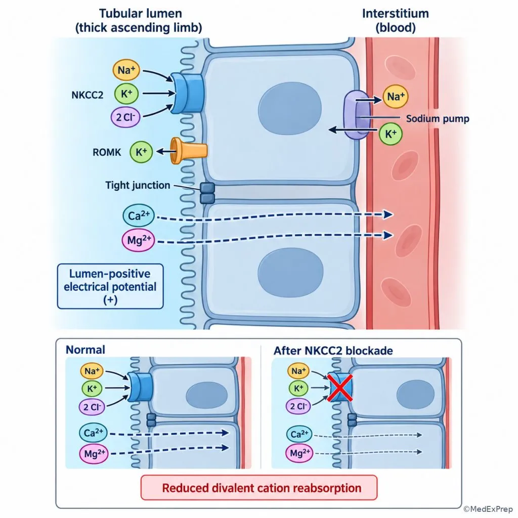
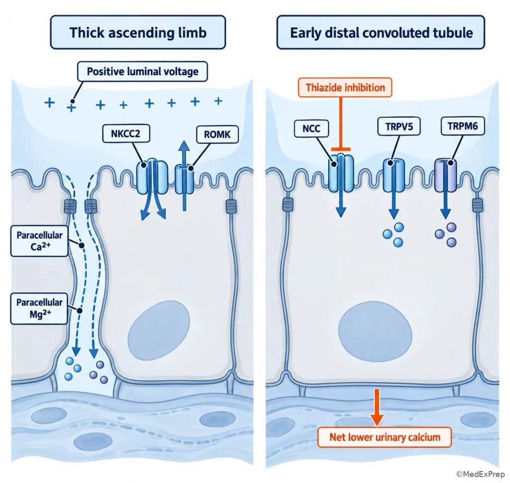
In the thick ascending limb, apical NKCC2 moves sodium, potassium, and chloride into tubular cells. ROMK recycles potassium into the lumen, supporting continued cotransporter function and a lumen-positive electrical potential. This favors paracellular calcium and magnesium reabsorption. The thick ascending limb has low water permeability and therefore dilutes tubular fluid while contributing to the medullary osmotic gradient.

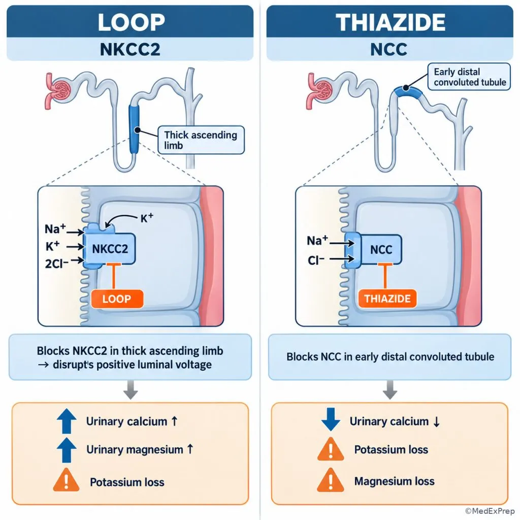
In the early distal convoluted tubule, NCC mediates sodium-chloride uptake. Distal calcium transport is regulated through pathways including TRPV5 and basolateral calcium extrusion; magnesium conservation depends in part on TRPM6. Thiazide-mediated NCC inhibition generally lowers urinary calcium excretion, whereas loop-mediated NKCC2 inhibition increases calcium and magnesium excretion. Sodium delivery beyond both sites may promote potassium secretion in downstream segments and contribute to metabolic alkalosis in the appropriate clinical setting.

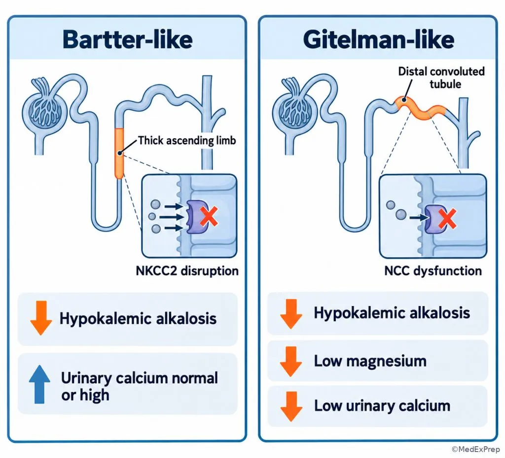
Decision rule: Choose NKCC2-related dysfunction when a salt-wasting pattern includes increased urinary calcium; favor an NCC-related pattern when hypocalciuria and hypomagnesemia accompany hypokalemic alkalosis, recognizing that clinical findings can vary.

See Fig 3 for the head-to-head transporter comparison.

Step 1 - Confirm pattern. Identify potassium, bicarbonate, magnesium, and blood pressure abnormalities.
Step 2 - Identify transporter exposure. Check loop and thiazide use, including possible unreported use.
Step 3 - Use calcium excretion. Increased urinary calcium suggests TAL dysfunction; reduced urinary calcium favors DCT NCC dysfunction.
Step 4 - Validate. Interpret urinary calcium with kidney function, sodium intake, volume state, and the clinical history.
Fig 3. Thick ascending limb versus early distal convoluted tubule
Exam takeaway: NKCC2 inhibition reduces the lumen-positive calcium-reabsorptive force, whereas NCC inhibition commonly reduces urinary calcium.
Electrochemical driving forces for divalent cations
Exam takeaway: Paracellular calcium and magnesium recovery in the thick ascending limb depends on the transport-generated electrical environment.

B. Worked example

A 68-year-old man with heart failure, hypertension, and chronic kidney disease develops muscle cramps after an increase in furosemide. Potassium is 3.1 mEq/L, magnesium is reduced, and serum bicarbonate is elevated. A recent urine collection suggests increased calcium excretion. Glucose is mildly elevated, and the patient has a remote history of gout.

  1. Identify the task: Determine the transporter most directly responsible for the coordinated electrolyte response.
  2. Extract key facts: Recent loop-diuretic exposure, hypokalemia, hypomagnesemia, alkalosis, and increased calcium excretion.
  3. Apply the rule: NKCC2 blockade disrupts thick ascending limb salt transport and its lumen-positive paracellular divalent cation gradient.
  4. Eliminate alternatives: NCC blockade can also cause hypokalemic alkalosis but typically reduces calcium excretion; mild hyperglycemia and prior gout do not explain the full tubular pattern.
  5. Verify: The direction of calcium and magnesium losses fits loop inhibition and warrants assessment of the dosing strategy, volume state, and electrolyte replacement needs.

Most likely mechanism: NKCC2 inhibition in the thick ascending limb.

C. Exam trap

Common wrong answer: Select NCC because thiazide-associated potassium depletion also causes alkalosis. Why it is tempting: Both classes increase downstream sodium delivery and can promote kaliuresis. Single eliminating clue: Increased urinary calcium points toward a loop effect rather than the usual thiazide effect.

D. Checkpoint MCQ

A normotensive adult has chronic hypokalemic metabolic alkalosis, hypomagnesemia, and low urinary calcium without diuretic exposure. Which transporter is most likely defective?

  1. A. NKCC2
  2. B. ENaC
  3. C. NCC
  4. D. SGLT2

Correct answer: C.

  • A: An NKCC2 defect can cause salt-wasting alkalosis, but the combination of hypocalciuria and magnesium depletion favors an NCC-related phenotype.
  • B: ENaC disturbance affects distal sodium and potassium handling, but isolated ENaC activation usually produces hypertension rather than this salt-wasting picture.
  • C: NCC loss of function produces the Gitelman-like combination of hypokalemic alkalosis, magnesium depletion, and reduced urinary calcium.
  • D: SGLT2 affects proximal glucose uptake and does not characteristically produce the specified distal magnesium-calcium phenotype.
If you missed this: Review Thick ascending limb and distal convoluted tubule: interpreting calcium and magnesium - focus on hypocalciuria as a clue favoring NCC dysfunction.
Quick Recall:

  • Complete the pair: Loop diuretic target = ____; thiazide target = ____. Answer: NKCC2; NCC.
  • True or false: Generalized proximal tubule failure can produce euglycemic glucosuria together with phosphate wasting. Answer: True.
  • One-liner: What helps distinguish Gitelman-like from Bartter-like physiology? Answer: Hypocalciuria with magnesium depletion favors Gitelman physiology.

3. Collecting duct: separate sodium, potassium, acid, and water regulation

A. Explanation

WHY THIS IS TESTED: Hyperkalemia, metabolic acidosis, and impaired water balance may all involve the distal nephron, but they are not interchangeable mechanisms. Board questions test whether the abnormality primarily reflects aldosterone-dependent electrolyte handling, intercalated-cell acid-base function, or vasopressin-dependent water permeability.

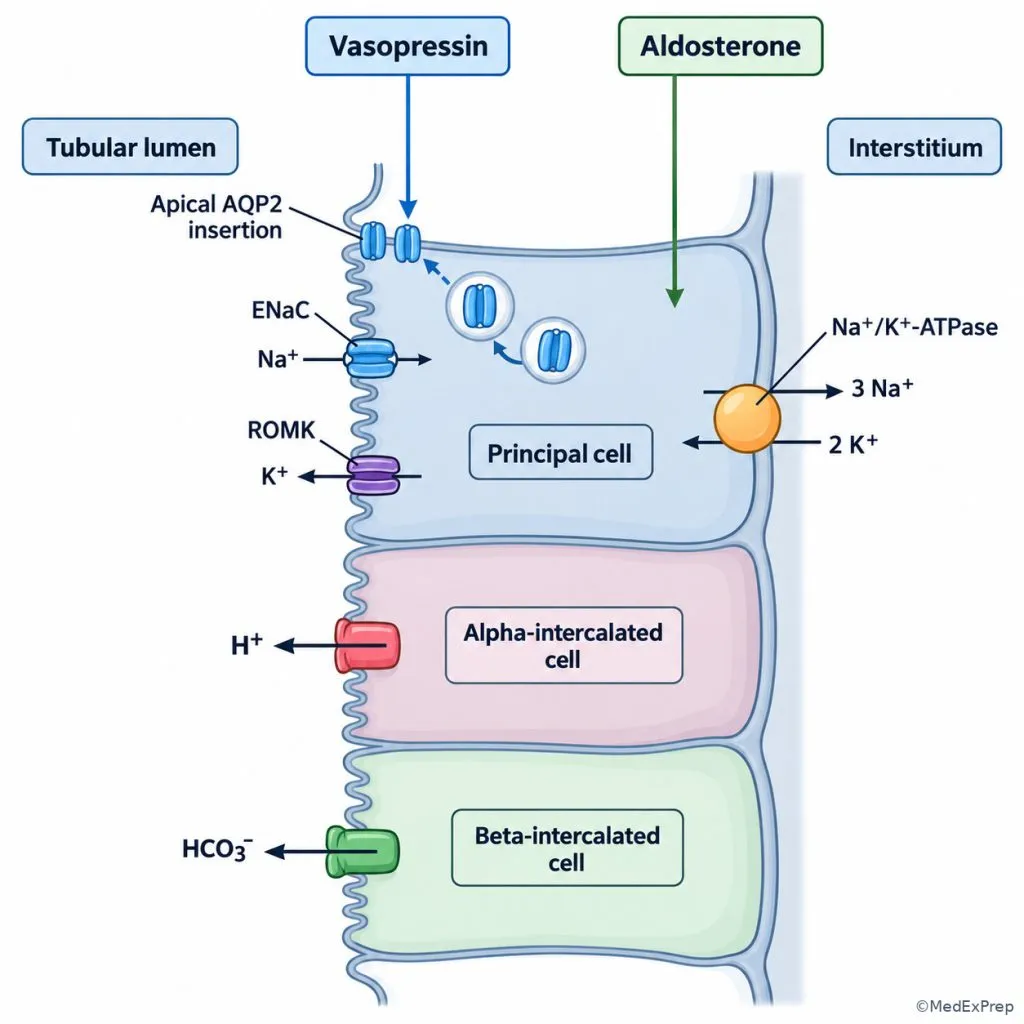
In principal cells of the aldosterone-sensitive distal nephron, ENaC allows electrogenic sodium entry. Basolateral Na+/K+-ATPase sustains the sodium gradient, while potassium channels allow potassium secretion. Aldosterone increases the capacity for sodium reabsorption and potassium secretion. The magnitude of potassium secretion also depends on distal sodium delivery, tubular flow, and the prevailing potassium load. Thus, hyperkalemia cannot be assigned to aldosterone deficiency solely from a single serum value.

Intercalated cells participate in urinary acidification and bicarbonate balance. Alpha-intercalated cells contribute to hydrogen secretion, whereas beta-intercalated cells support bicarbonate secretion. Reduced aldosterone effect, particularly in diabetes or chronic kidney disease, can produce hyperkalemia with a normal-anion-gap metabolic acidosis, conventionally termed type 4 renal tubular acidosis. Reduced ammonium production and excretion are important contributors; urine may still become appropriately acidic, so urine pH alone does not exclude this syndrome.

Vasopressin works through V2-mediated AQP2 recruitment in principal cells, increasing collecting duct water permeability. It regulates water recovery rather than directly substituting for aldosterone-mediated potassium control. The medullary gradient, tubular delivery, and vasopressin response together determine concentrating ability; this is developed further in Urine Concentration And Dilution.

Decision rule: Interpret hyperkalemia through aldosterone action and distal delivery; interpret disproportionate water retention through collecting duct water permeability. See Fig 4.

Step 1 - Identify the dominant disturbance. Is the clinical problem potassium, acid-base, sodium balance, or free-water regulation?
Step 2 - Check distal sodium handling. Evaluate effective circulating volume, kidney function, and medication effects on distal delivery.
Step 3 - Evaluate hormones. Distinguish aldosterone-mediated sodium-potassium control from vasopressin-mediated water permeability.
Step 4 - Integrate acid-base data. Hyperkalemia with normal-gap acidosis suggests reduced ammonium excretion or aldosterone effect in the appropriate setting.
Fig 4. Distal nephron potassium reasoning
Exam takeaway: Potassium concentration must be interpreted with acid-base status, distal sodium delivery, and mineralocorticoid action.
flowchart TD
A[Potassium abnormality] --> B{High or low potassium}
B -->|High| C[Assess kidney function and distal delivery]
C --> D[Review RAAS drugs and aldosterone action]
D --> E[Check for normal-gap acidosis]
B -->|Low| F[Assess renal losses and diuretic exposure]
F --> G[Use blood pressure and acid-base pattern]
G --> H[Consider ENaC activation or increased distal delivery]
Principal and intercalated cell transport
Exam takeaway: The aldosterone-sensitive sodium-potassium pathway and intercalated-cell acid-base pathways are functionally distinct.

B. Worked example

A 64-year-old man with type 2 diabetes, hypertension, and moderately reduced chronic kidney function has potassium 5.8 mEq/L and bicarbonate 19 mEq/L. Sodium is 140 mEq/L and chloride is 111 mEq/L, giving an anion gap of 10 mEq/L. Kidney function is near baseline. He takes an ACE inhibitor for albuminuric kidney disease. Urine pH is 5.2, electrocardiography shows no acute hyperkalemic changes, and trace proteinuria is present.

  1. Identify the task: Explain hyperkalemia with normal-anion-gap acidosis despite relatively stable filtration.
  2. Extract key facts: Diabetes, RAAS blockade, potassium elevation, hyperchloremic acidosis, and no major acute GFR decline.
  3. Apply the rule: Reduced effective aldosterone signaling can impair potassium excretion and ammonium-mediated net acid excretion.
  4. Eliminate alternatives: Distal type 1 RTA more often presents with impaired urine acidification and low potassium; a urine pH below 5.5 does not eliminate type 4 physiology.
  5. Verify: Review medications, volume status, dietary intake, kidney function, and the severity of hyperkalemia; choose potassium-directed treatment and consider changes to RAAS therapy according to clinical risk and benefit.

Most likely physiology: Reduced mineralocorticoid effectiveness with a type 4 RTA pattern. A normal electrocardiogram does not establish that hyperkalemia is harmless.

C. Exam trap

Common wrong answer: Exclude renal tubular acidosis because urine pH is acidic. Why it is tempting: Failure to lower urine pH is a familiar discriminator for classic distal type 1 RTA. Single eliminating clue: The combination of hyperkalemia and normal-gap acidosis is compatible with reduced ammonium excretion in type 4 RTA even when urine pH is low.

D. Checkpoint MCQ

Which finding most strongly supports impaired aldosterone action rather than isolated vasopressin deficiency?

  1. A. Polyuria with dilute urine and normal serum potassium
  2. B. Hyperkalemia with normal-anion-gap metabolic acidosis
  3. C. Hypernatremia with low urine osmolality
  4. D. Impaired concentrating ability after loss of the medullary gradient

Correct answer: B.

  • A: Polyuria is tempting because both hormones act in the distal nephron, but dilute urine points toward water permeability or concentrating mechanisms.
  • B: Potassium retention with hyperchloremic acidosis is the characteristic electrolyte-acid-base signature of reduced mineralocorticoid effectiveness.
  • C: Hypernatremia with dilute urine raises concern for water loss and vasopressin-related physiology rather than a primary aldosterone effect.
  • D: Medullary gradient disruption affects urinary concentration but does not specifically identify defective mineralocorticoid signaling.
If you missed this: Review Collecting duct: separate sodium, potassium, acid, and water regulation - focus on aldosterone governing distal potassium handling and vasopressin governing water permeability.

4. Diuretics: transporter inhibition as a physiologic experiment

A. Explanation

WHY THIS IS TESTED: A stem may ask for the best diuretic class, the most likely adverse electrolyte change, or the transporter affected by a medication. Recognizing the nephron location is more reliable than memorizing drugs as isolated facts. Drug selection still depends on the therapeutic goal, comorbidities, filtration capacity, volume state, and laboratory values.

Carbonic anhydrase inhibitors impair proximal bicarbonate recovery and can cause bicarbonaturia and metabolic acidosis. SGLT2 inhibitors reduce proximal glucose reabsorption, producing glycosuria and associated osmotic effects. Loop agents block NKCC2, strongly affecting salt reabsorption and increasing urinary calcium and magnesium loss. Thiazide and thiazide-like agents block NCC, reducing urinary calcium in typical circumstances. ENaC blockers and mineralocorticoid receptor antagonists decrease distal potassium secretion and can precipitate hyperkalemia, especially with reduced GFR or concurrent RAAS blockade.

Both loop and thiazide classes may increase potassium loss because sodium reaching downstream electrogenic sodium-reabsorbing sites can enhance potassium secretion. Potassium-sparing agents oppose this effect by decreasing the driving force or hormonal stimulus for secretion. The term potassium-sparing does not imply safety in a patient with existing hyperkalemia.

Decision rule: Match the therapeutic objective to the transporter, then screen the predicted electrolyte consequence before selecting a medication. See Fig 5 for the high-yield loop versus thiazide decision.

Step 1 - Define the goal. Determine whether treatment targets extracellular fluid volume, blood pressure, calcium excretion, bicarbonate handling, or potassium conservation.
Step 2 - Identify transporter site. Locate the intended drug effect on proximal, TAL, DCT, or collecting duct transport.
Step 3 - Predict accompanying changes. Anticipate sodium, potassium, calcium, magnesium, bicarbonate, and water effects.
Step 4 - Screen contraindications and interactions. Consider kidney function, volume depletion, baseline potassium, blood pressure, and other medications.
Step 5 - Monitor. Reassess symptoms, hemodynamics, kidney function, and electrolyte trends after treatment changes.
Table 2. Diuretic classes and characteristic transport consequences
Class Primary target Expected effect High-yield safety clue
Carbonic anhydrase inhibitor Proximal bicarbonate reclamation Increased bicarbonate excretion Can produce or worsen metabolic acidosis.
SGLT2 inhibitor Proximal sodium-glucose cotransport Glucosuria and osmotic effects Glucosuria is not by itself evidence of Fanconi syndrome.
Loop diuretic NKCC2, thick ascending limb Increased sodium, calcium, and magnesium excretion; possible potassium loss Electrolyte depletion and volume contraction require monitoring.
Thiazide or thiazide-like diuretic NCC, early DCT Increased NaCl excretion with typically reduced urinary calcium Monitor potassium, sodium, and effects on metabolic comorbidities.
ENaC inhibitor ENaC, aldosterone-sensitive distal nephron Reduced sodium uptake and potassium secretion Hyperkalemia risk increases with CKD and RAAS blockade.
Mineralocorticoid receptor antagonist Aldosterone signaling Reduced sodium retention and potassium secretion Assess potassium and kidney function before and after initiation.
Fig 5. Loop versus thiazide decision comparison
Exam takeaway: Urinary calcium increases with loop inhibition but commonly decreases with thiazide inhibition.

B. Worked example

A 58-year-old woman with hypertension, recurrent calcium-containing kidney stones, gout, and mildly reduced kidney function has increased urinary calcium on a metabolic stone evaluation. She is not volume overloaded. Her potassium is normal, and serum uric acid is mildly elevated. Her clinician is considering a blood pressure medication that may also reduce urinary calcium; sodium reduction and fluid-intake counseling are being addressed.

  1. Identify the task: Select the transporter target most likely to lower urinary calcium while treating hypertension.
  2. Extract key facts: Hypercalciuria, hypertension, no compelling volume-overload indication for loop therapy, and gout as a treatment limitation.
  3. Apply the rule: NCC inhibition generally decreases urinary calcium excretion and may be considered when the overall clinical indication favors a thiazide-type agent.
  4. Eliminate alternatives: Loop inhibition tends to increase urinary calcium; ENaC blockade and carbonic anhydrase inhibition do not provide the same characteristic calcium effect.
  5. Verify: Consider gout risk, sodium and potassium effects, kidney function, blood pressure, and the benefits of dietary sodium reduction before selecting and monitoring treatment.

Best transporter target for reducing urinary calcium: NCC. This mechanistic preference does not automatically establish that a thiazide is the best overall medication for every patient with stones and gout.

C. Exam trap

Common wrong answer: Choose a loop diuretic because loops and thiazides both increase urine output. Why it is tempting: Both inhibit sodium reabsorption and can cause potassium depletion. Single eliminating clue: The stated goal is reduced urinary calcium, which favors NCC rather than NKCC2 inhibition.

D. Checkpoint MCQ

Which medication mechanism is most directly associated with a decrease in urinary calcium excretion?

  1. A. Blockade of NKCC2
  2. B. Blockade of NCC
  3. C. Inhibition of proximal carbonic anhydrase
  4. D. Antagonism of vasopressin V2 receptors

Correct answer: B.

  • A: NKCC2 blockade is tempting because it is an effective natriuretic mechanism, but it disrupts TAL calcium reabsorption and increases urinary calcium.
  • B: NCC inhibition characteristically increases net distal calcium conservation and reduces calcium excretion.
  • C: Carbonic anhydrase inhibition causes bicarbonaturia, but this is not the standard mechanism for reducing urinary calcium.
  • D: V2 antagonism increases electrolyte-free water excretion rather than selectively promoting calcium conservation.
If you missed this: Review Diuretics: transporter inhibition as a physiologic experiment - focus on the opposite urinary calcium responses to loop and thiazide inhibition.
Quick Recall:

  • Fill in the blank: Amiloride reduces potassium secretion by blocking ____. Answer: ENaC.
  • True or false: A potassium-sparing diuretic is automatically safe when baseline potassium is elevated. Answer: False.
  • One-liner: Which hormone changes collecting duct water permeability through AQP2? Answer: Vasopressin.

5. Inherited transport disorders: phenotype-to-segment localization

A. Explanation

WHY THIS IS TESTED: Inherited tubular syndromes can resemble chronic exposure to a particular diuretic. The board-relevant challenge is to discriminate disorders with similar potassium and bicarbonate findings by incorporating blood pressure, calcium excretion, magnesium, renin, aldosterone, and the suspected transporter.

Bartter syndromes affect transport pathways supporting thick ascending limb salt reabsorption, including NKCC2, ROMK, and related channels or proteins. Patients typically demonstrate a salt-wasting, hypokalemic metabolic alkalosis phenotype; urinary calcium may be normal or increased depending on subtype and circumstances. Gitelman syndrome reflects NCC dysfunction and typically produces hypokalemic alkalosis, hypomagnesemia, and hypocalciuria. A loop-like versus thiazide-like physiologic pattern is more discriminating than age at presentation alone.

Liddle syndrome results from increased ENaC activity and produces inappropriate distal sodium retention, hypertension, often hypokalemia and alkalosis, and suppressed renin and aldosterone. Since the problem lies in the channel rather than aldosterone excess, direct ENaC blockade is mechanistically appropriate. Gordon syndrome, also called pseudohypoaldosteronism type II, features increased NCC-mediated sodium chloride transport due to regulatory pathway abnormalities and is associated with hypertension, hyperkalemia, and often metabolic acidosis; thiazide sensitivity is an important clue. The terms pseudohypoaldosteronism type I and type II describe different physiologic syndromes and should not be conflated.

Fanconi syndrome belongs in the differential of a different pattern: generalized proximal solute wasting rather than a predominantly salt-wasting distal alkalosis syndrome. Some inherited disorders cause Fanconi physiology, but acquired causes such as medications must also be considered in adults.

Decision rule: Begin with blood pressure and potassium, then use acid-base status, magnesium, calcium excretion, and renin-aldosterone patterns to identify the likely transporter. A phenotype suggests the pathway; genetics or specialized testing may be required to establish a molecular diagnosis.

Step 1 - Separate hypertensive from salt-wasting phenotypes. Blood pressure identifies whether sodium retention or loss predominates.
Step 2 - Classify potassium and bicarbonate. Identify hypokalemic alkalosis versus hyperkalemic acidosis.
Step 3 - Examine calcium and magnesium. Distinguish a Gitelman-like NCC pattern from a Bartter-like loop pattern.
Step 4 - Interpret renin and aldosterone. Identify suppressed hormonal signaling with inappropriate channel activation versus true mineralocorticoid excess.
Step 5 - Confirm clinically. Exclude diuretic use and acquired causes before selecting genetic testing or transporter-directed treatment.
Table 3. Major inherited and acquired tubular transport phenotypes
Phenotype Primary pathway Typical laboratory or clinical signature Key distinction
Fanconi-type proximal dysfunction Multiple proximal reabsorptive processes Glucosuria without hyperglycemia, phosphate wasting, bicarbonate loss and possibly other proximal losses Multisubstrate wasting, not isolated distal potassium loss.
Bartter syndrome TAL salt-transport pathways Salt wasting, hypokalemic alkalosis, often increased renin and aldosterone; calcium excretion variable Loop-like physiology without loop exposure.
Gitelman syndrome NCC loss of function Hypokalemic alkalosis, low magnesium, low urinary calcium Thiazide-like physiology without thiazide exposure.
Liddle syndrome ENaC gain of function Hypertension, frequently hypokalemia and alkalosis, low renin and low aldosterone Direct ENaC overactivity rather than aldosterone excess.
Gordon syndrome Increased NCC pathway activity Hypertension, hyperkalemia, frequently normal-gap acidosis Hyperkalemic hypertension with thiazide-responsive physiology.
Reduced aldosterone effect or type 4 RTA Mineralocorticoid signaling and ammonium-excretion pathways Hyperkalemia with normal-gap metabolic acidosis Common acquired context includes diabetes, CKD, and medication effects.
Blood pressure and potassium phenotype decision tree
Exam takeaway: Hypertension with low potassium suggests sodium-retaining ENaC activity, whereas hypertension with high potassium raises an NCC regulatory or aldosterone-related differential.
flowchart TD
A[Suspected tubular transport syndrome] --> B{Hypertension present}
B -->|No| C{Hypokalemic alkalosis}
C -->|Yes| D[Evaluate magnesium and urinary calcium]
D --> E[Gitelman-like or Bartter-like]
B -->|Yes| F{Potassium low or high}
F -->|Low| G[Assess renin and aldosterone for Liddle pattern]
F -->|High| H[Assess CKD drugs and Gordon pattern]
C -->|No| I[Consider proximal solute wasting or alternative disorder]
Gitelman-like versus Bartter-like electrolyte signatures
Exam takeaway: Hypomagnesemia with hypocalciuria favors the NCC-related Gitelman phenotype over a typical loop-like salt-wasting pattern.
Hypertensive tubular disorders comparison
Exam takeaway: Liddle syndrome usually produces hypokalemic alkalosis, while Gordon syndrome produces hyperkalemia and often acidosis despite hypertension in both.

B. Worked example

A 41-year-old woman with hypothyroidism, asthma, and migraines is evaluated for recurrent muscle cramps. She is not taking diuretics. Blood pressure is 106/68 mmHg. Potassium is 2.9 mEq/L, bicarbonate is 32 mEq/L, and magnesium is low. Urinary calcium is low. A mildly reduced serum phosphate level is noted, but there is no glucosuria or other evidence of generalized proximal wasting.

  1. Identify the task: Determine which inherited transporter phenotype best explains chronic hypokalemic alkalosis.
  2. Extract key facts: Low-normal blood pressure, hypomagnesemia, hypocalciuria, and absence of diuretic use.
  3. Apply the rule: The constellation is most characteristic of impaired NCC activity in the distal convoluted tubule.
  4. Eliminate alternatives: Bartter syndrome may cause alkalosis but is less strongly associated with the hypocalciuric-magnesium pattern; Liddle syndrome typically causes hypertension; mild isolated phosphate reduction is insufficient for Fanconi syndrome.
  5. Verify: Reassess medication exposure, urinary electrolyte findings, family history, and appropriate confirmatory testing while managing magnesium and potassium depletion.

Most likely diagnosis: Gitelman syndrome or a Gitelman-like NCC transport phenotype.

C. Exam trap

Common wrong answer: Diagnose Bartter syndrome whenever hypokalemia and metabolic alkalosis coexist. Why it is tempting: Both Bartter and Gitelman syndromes produce salt-wasting activation of downstream potassium-secretory mechanisms. Single eliminating clue: Marked hypocalciuria with hypomagnesemia strongly favors Gitelman physiology.

D. Checkpoint MCQ

An adult has early-onset hypertension, potassium 2.8 mEq/L, metabolic alkalosis, suppressed renin, and suppressed aldosterone. Which primary mechanism best explains the presentation?

  1. A. Autonomous ENaC activity
  2. B. Increased adrenal aldosterone secretion
  3. C. Loss of NCC activity
  4. D. Loss of NKCC2 activity

Correct answer: A.

  • A: ENaC gain of function causes sodium retention and potassium secretion without requiring elevated aldosterone, matching the Liddle pattern.
  • B: Aldosterone excess is tempting because it also causes hypertension with hypokalemic alkalosis, but autonomous aldosterone production would not typically suppress aldosterone itself.
  • C: NCC loss produces salt wasting and generally low or normal blood pressure rather than the hypertensive sodium-retaining picture.
  • D: NKCC2 loss is a loop-like salt-wasting mechanism with secondary RAAS activation, inconsistent with the suppressed renin-aldosterone signature.
If you missed this: Review Inherited transport disorders: phenotype-to-segment localization - focus on low renin plus low aldosterone in hypokalemic hypertension as the Liddle discriminator.

Exam Traps & Differentiators

Strong distractors often preserve part of the correct physiology. The discriminating task is to find the one finding that cannot be explained by the tempting alternative.

Common traps & distractors:

  • Glucosuria means uncontrolled diabetes. Tempting because hyperglycemia increases filtered glucose; eliminating clue: glucosuria persists with normal plasma glucose.
  • Any euglycemic glucosuria is Fanconi syndrome. Tempting because Fanconi affects proximal glucose recovery; eliminating clue: isolated glucosuria without other proximal losses is insufficient.
  • Loop and thiazide agents have identical electrolyte effects. Tempting because both increase natriuresis and may lower potassium; eliminating clue: their usual urinary calcium directions differ.
  • Acidic urine excludes type 4 RTA. Tempting because classic distal RTA is associated with impaired acidification; eliminating clue: type 4 RTA primarily features hyperkalemia with reduced ammonium-mediated acid excretion.
  • Low aldosterone excludes a mineralocorticoid-like syndrome. Tempting because mineralocorticoid excess causes sodium retention; eliminating clue: Liddle syndrome activates ENaC independently of aldosterone.
  • All hypokalemic alkalosis indicates the same inherited disease. Tempting because loop and distal salt-wasting mechanisms converge downstream; eliminating clue: hypomagnesemia and hypocalciuria favor Gitelman physiology.
Table 4. Looks similar but is not: critical nephrology differentials
First possibility Tempting alternative Decisive distinction
Generalized proximal solute wasting Isolated SGLT2-related glucosuria Additional inappropriate phosphate, bicarbonate, or other proximal substrate losses.
Bartter-like defect Gitelman-like defect Urinary calcium and magnesium patterns help discriminate the involved segment.
Loop-diuretic effect Thiazide effect Loop blockade promotes calciuria; thiazide blockade usually reduces it.
Liddle syndrome Primary aldosteronism Both can cause hypokalemic hypertension, but aldosterone is suppressed in Liddle and inappropriately elevated in primary aldosteronism.
Gordon syndrome Acquired type 4 RTA Both may produce hyperkalemic acidosis; hypertension, lifelong history, drug exposure, kidney function, and NCC-related phenotype guide evaluation.
Vasopressin-related water-handling defect Mineralocorticoid-related potassium defect Disproportionate dilute polyuria points toward water handling; hyperkalemia with normal-gap acidosis points toward aldosterone-dependent physiology.

If the stem says, think

  • Euglycemic glucosuria plus phosphaturia: generalized proximal dysfunction.
  • Loop exposure with magnesium loss and calciuria: disrupted thick ascending limb transport.
  • Hypokalemic alkalosis plus hypocalciuria and low magnesium: NCC loss-of-function pattern.
  • Hypertension, hypokalemia, low renin, low aldosterone: ENaC gain-of-function pattern.
  • Diabetes or RAAS blockade plus hyperkalemic normal-gap acidosis: impaired aldosterone effectiveness or type 4 RTA physiology.
  • Hypertension, hyperkalemia, acidosis, and a thiazide-sensitive inherited phenotype: Gordon syndrome.
Review connection: Return to Inherited transport disorders: phenotype-to-segment localization when both potassium and blood pressure are abnormal; return to Thick ascending limb and distal convoluted tubule when calcium or magnesium excretion separates two otherwise similar diagnoses.

Algorithm / Approach

Five-step segmental localization framework: Apply the same sequence to an unexplained electrolyte disturbance, medication adverse effect, or suspected inherited tubulopathy.

1. Identify the task. Determine whether the question asks for a segment, transporter, syndrome, medication effect, or best management action.
2. Extract the pattern. Organize serum potassium, bicarbonate, magnesium, phosphate, glucose, urinary calcium, blood pressure, and kidney function.
3. Apply segment physiology. Proximal: multiple solutes; TAL: salt and divalent cation gradients; DCT: NCC, calcium and magnesium; collecting duct: potassium, acid, and water regulation.
4. Eliminate mimics. Review diuretics, SGLT2 inhibitors, RAAS medications, endocrine effects, volume state, and acquired kidney injury before labeling a genetic defect.
5. Verify and act. Reconcile all major abnormalities, address urgent electrolyte danger, select focused testing, and make a kidney-appropriate treatment decision.

Safety screen: Severe or symptomatic potassium disturbances, concerning electrocardiographic changes, significant acid-base instability, or substantial volume abnormalities require timely treatment and escalation while the underlying transporter mechanism is investigated. Local protocols vary; confirm with your institution.

Self-check quiz

Answer these five independent board-style questions before opening the explanations. Each asks for the best single answer, not merely a plausible association.

Question 1 - Foundational localization

A 60-year-old patient treated with a potentially proximal-toxic medication has normal serum glucose but glucosuria, hypophosphatemia with renal phosphate loss, and normal-anion-gap metabolic acidosis. Which process best integrates these findings?

  1. A. Reduced NKCC2 transport in the thick ascending limb
  2. B. Reduced NCC transport in the early distal tubule
  3. C. Generalized proximal tubular reabsorptive dysfunction
  4. D. Selective impairment of collecting duct potassium secretion

Question 2 - Mechanism of calciuria

A patient with edema receives an intravenous loop diuretic and subsequently develops increased urinary calcium and magnesium excretion. Which immediate change best explains this?

  1. A. Inhibition of proximal sodium-phosphate cotransport
  2. B. Reduced NKCC2-mediated transport and loss of lumen-positive potential
  3. C. Increased NCC-dependent distal calcium conservation
  4. D. Inhibition of ENaC-driven electrogenic sodium entry

Question 3 - Integrative pharmacology

A 36-year-old patient with hypertension, hypokalemic metabolic alkalosis, low renin, and low aldosterone is diagnosed with Liddle syndrome. Which therapy most directly addresses the abnormal transporter?

  1. A. Spironolactone
  2. B. Amiloride
  3. C. Hydrochlorothiazide
  4. D. Acetazolamide

Question 4 - Acid-base interpretation

A 67-year-old patient with diabetic kidney disease has potassium 5.7 mEq/L, bicarbonate 18 mEq/L, a normal anion gap, stable kidney function, and urine pH 5.1. Which mechanism is most consistent with the pattern?

  1. A. Isolated loss of collecting duct water permeability
  2. B. Isolated distal hydrogen secretion failure with typical hypokalemia
  3. C. Increased thiazide-sensitive NaCl reabsorption with obligatory hypokalemia
  4. D. Reduced aldosterone effectiveness with impaired ammonium-mediated acid excretion

Question 5 - Segment-specific decision

An adult with hypertension and recurrent calcium stones has documented hypercalciuria. Assuming a diuretic is otherwise clinically appropriate, which inhibition is most likely to reduce urinary calcium?

  1. A. NKCC2 inhibition
  2. B. NCC inhibition
  3. C. Carbonic anhydrase inhibition
  4. D. Vasopressin V2 receptor inhibition

Answer key & explanations

Question 1: C - Generalized proximal tubular reabsorptive dysfunction

The critical finding is loss of more than one normally reclaimed proximal solute. The pattern is compatible with Fanconi-type physiology; investigation must distinguish causes, including medications.

  • A - Incorrect: TAL salt transport failure is tempting because it can disturb electrolytes and acid-base balance, but it does not account for simultaneous euglycemic glucosuria and phosphaturia.
  • B - Incorrect: NCC disorders affect sodium, magnesium, calcium, and downstream potassium handling, but do not explain glucose and phosphate wasting together.
  • C - Correct: Proximal dysfunction links the glucose, phosphate, and bicarbonate abnormalities through reduced reabsorption of several substrates.
  • D - Incorrect: Impaired distal potassium secretion can produce hyperkalemia and acidosis, but cannot explain the combination of glucosuria and renal phosphate wasting.
If you missed this: Review Proximal tubule: bulk recovery versus generalized solute wasting - focus on recognizing multisubstrate proximal reabsorption failure.

Question 2: B - Reduced NKCC2 transport and lumen-positive potential

Loop inhibition interferes with TAL sodium-potassium-chloride uptake and the associated voltage that normally favors paracellular calcium and magnesium reabsorption.

  • A - Incorrect: Proximal phosphate cotransport affects phosphate balance and may appear relevant to mineral disorders, but it is not the immediate mechanism of loop-associated calcium and magnesium loss.
  • B - Correct: Reduced NKCC2 function and potassium recycling diminish the lumen-positive driving force, increasing divalent cation excretion.
  • C - Incorrect: NCC-associated distal calcium conservation is a tempting calcium-related mechanism, but it describes a thiazide-associated direction rather than loop-associated calciuria.
  • D - Incorrect: ENaC affects electrogenic sodium and potassium transport downstream, but does not create the TAL calcium-magnesium gradient disrupted by loop diuretics.
If you missed this: Review Thick ascending limb and distal convoluted tubule: interpreting calcium and magnesium - focus on the NKCC2-ROMK voltage mechanism.

Question 3: B - Amiloride

Liddle syndrome is an ENaC gain-of-function disorder. Direct channel inhibition addresses inappropriate sodium reabsorption and downstream potassium secretion despite suppressed aldosterone.

  • A - Incorrect: Spironolactone is tempting because mineralocorticoid excess can also cause hypokalemic hypertension, but Liddle ENaC activity is not driven by excessive aldosterone receptor stimulation.
  • B - Correct: Amiloride directly inhibits ENaC, targeting the transporter responsible for the syndrome.
  • C - Incorrect: Hydrochlorothiazide changes upstream NCC activity and can lower blood pressure, but it does not specifically reverse the channel gain of function and may aggravate potassium depletion.
  • D - Incorrect: Acetazolamide modifies proximal bicarbonate recovery and acid-base handling, but it does not selectively inhibit inappropriate distal ENaC activity.
If you missed this: Review Inherited transport disorders: phenotype-to-segment localization - focus on ENaC gain of function with low renin and low aldosterone.

Question 4: D - Reduced aldosterone effectiveness and ammonium excretion

Hyperkalemia together with normal-anion-gap metabolic acidosis in diabetic kidney disease supports type 4 RTA physiology. A low urinary pH does not exclude this mechanism because ammonium generation and excretion, rather than free hydrogen ion concentration alone, are central to net acid elimination.

  • A - Incorrect: Collecting duct water-permeability abnormalities are tempting because they affect the same broad nephron region, but they principally alter water balance and urine concentration rather than causing this potassium-acid-base combination.
  • B - Incorrect: Classic distal type 1 RTA is a reasonable alternative for normal-gap acidosis, but typically involves impaired urine acidification and often hypokalemia, not this characteristic hyperkalemic pattern.
  • C - Incorrect: Increased NCC activity is tempting because Gordon syndrome can produce hypertension, hyperkalemia, and acidosis, but the proposed obligatory hypokalemia contradicts that syndrome and the wording of the option.
  • D - Correct: Reduced effective aldosterone signaling can impair potassium elimination and ammonium-mediated renal acid excretion, matching type 4 physiology.
If you missed this: Review Collecting duct: separate sodium, potassium, acid, and water regulation - focus on hyperkalemic normal-gap acidosis despite an appropriately acidic urine pH.

Question 5: B - NCC inhibition

Thiazide-type NCC inhibition typically decreases urinary calcium excretion, an effect potentially useful when hypertension and hypercalciuria coexist. The complete treatment decision also considers sodium intake, prior stone events, comorbidities, and medication tolerability.

  • A - Incorrect: NKCC2 blockade is tempting because it is a potent diuretic mechanism, but it tends to increase urinary calcium loss.
  • B - Correct: NCC inhibition commonly increases net renal calcium conservation and reduces urinary calcium excretion.
  • C - Incorrect: Carbonic anhydrase inhibition is tempting because proximal transport affects mineral and acid-base handling, but bicarbonaturia and altered urine chemistry do not make it the preferred calcium-conserving mechanism.
  • D - Incorrect: V2 receptor inhibition increases water excretion, which may change urine concentration, but it does not directly provide the characteristic reduction in calcium excretion sought here.
If you missed this: Review Diuretics: transporter inhibition as a physiologic experiment - focus on the opposite directions of urinary calcium handling during NCC and NKCC2 inhibition.

Rapid review / cheat-sheet

  • Proximal injury -> combined wasting of filtered substrates, not one isolated urinary abnormality.
  • Proximal bicarbonate -> NHE3 and carbonic anhydrase-dependent reclamation.
  • Phosphate regulation -> PTH and FGF23 decrease proximal phosphate reabsorption.
  • TAL NKCC2 -> salt uptake linked to a calcium-magnesium reabsorptive voltage.
  • Loop blockade -> more urinary calcium and magnesium.
  • DCT NCC -> thiazide-sensitive NaCl uptake and typically lower calcium excretion when inhibited.
  • Gitelman phenotype -> low magnesium plus low urinary calcium in hypokalemic alkalosis.
  • Principal cell ENaC -> sodium entry coupled to potassium-secretory forces.
  • Aldosterone impairment -> hyperkalemia and potentially normal-gap acidosis.
  • Vasopressin-AQP2 -> water permeability, not a substitute for aldosterone.
  • Liddle syndrome -> low-renin, low-aldosterone hypokalemic hypertension; directly target ENaC.
  • Gordon syndrome -> hyperkalemic hypertension with NCC regulatory overactivity and thiazide sensitivity.

See also

Prior foundation: GFR And Renal Blood Flow (ch01.s01.t01) establishes filtration and delivery. Next: Urine Concentration And Dilution (ch01.s01.t03) develops the water-handling consequences of the medullary gradient and vasopressin. Diagnostic extensions: Urine Sediment Patterns (ch01.s02.t01), Proteinuria And Albuminuria Assessment (ch01.s02.t02), and Fractional Excretion Reasoning (ch01.s02.t03). Clinical extensions: Hypokalemia Evaluation (ch02.s02.t02), Metabolic Acidosis Differentials (ch02.s03.t02), and Inherited Tubulopathies And Alport (ch06.s04.t02).

Blueprint anchor recap: PHYSIOLOGY_PATHOLOGY_DIAGNOSTICS -> renal-physiology-and-diagnostics -> proximal and distal transport mechanisms; FLUID_ELECTROLYTE_ACID_BASE -> segmental sodium, potassium, calcium, magnesium, phosphate, bicarbonate, and water interpretation. This lesson supplies a topic-level subskill map and makes no claim about undisclosed ABIM item weights.

Further study within the course: Renal Physiology, Glomerular And Tubular Physiology; Fluid Electrolyte Acid-Base, Potassium And Related Electrolytes; Tubulointerstitial Hypertension Stone Cystic, Inherited And Cystic Disorders. Detailed reference editions and page-specific citations were not supplied in the source pack.

Completion standard: Given an adult clinical vignette, identify the most likely nephron segment, support localization using two independent findings, exclude a plausible medication or endocrine mimic, and explain the expected consequence of transporter-directed treatment.

Keep reading, free

Enter your email to read lesson 2 now.

Free. We may also email you about our programs. No spam, and you can ask us to remove your email at any time.

Liked it?

ABIM Nephrology Prep Course (Nephrology) has 76 lessons.

ABIMPrep
Programs
For organizations
ContactLog inGet a free guide